. 2007 Nov;13(5):635-645.
doi: 10.1016/j.devcel.2007.10.006.

Insights into COPII coat nucleation from the structure of Sec23.Sar1 complexed with the active fragment of Sec31

Affiliations

Insights into COPII coat nucleation from the structure of Sec23.Sar1 complexed with the active fragment of Sec31

Xiping Bi et al. Dev Cell. 2007 Nov.

Abstract

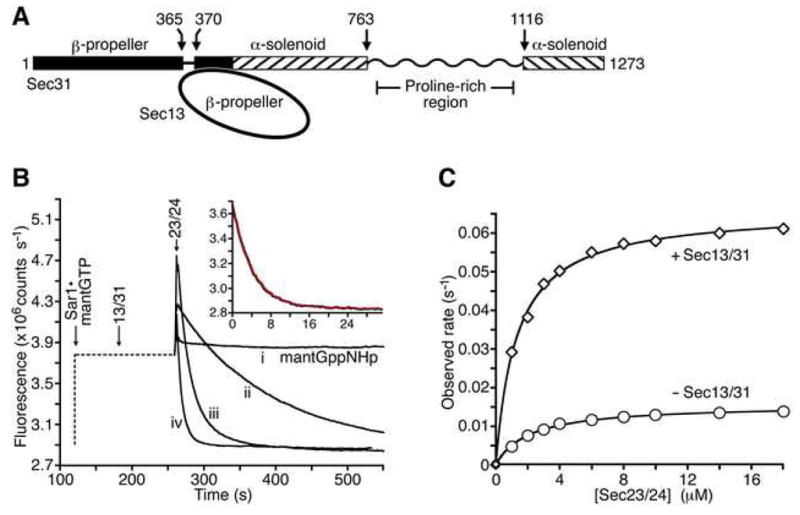
The COPII vesicular coat forms on the endoplasmic reticulum from Sar1-GTP, Sec23/24 and Sec13/31 protein subunits. Here, we define the interaction between Sec23/24.Sar1 and Sec13/31, involving a 40 residue Sec31 fragment. In the crystal structure of the ternary complex, Sec31 binds as an extended polypeptide across a composite surface of the Sec23 and Sar1-GTP molecules, explaining the stepwise character of Sec23/24.Sar1 and Sec13/31 recruitment to the membrane. The Sec31 fragment stimulates GAP activity of Sec23/24, and a convergence of Sec31 and Sec23 residues at the Sar1 GTPase active site explains how GTP hydrolysis is triggered leading to COPII coat disassembly. The Sec31 active fragment is accommodated in a binding groove supported in part by Sec23 residue Phe380. Substitution of the corresponding residue F382L in human Sec23A causes cranio-lenticulo-sutural dysplasia, and we suggest that this mutation disrupts the nucleation of COPII coat proteins at endoplasmic reticulum exit sites.


Figures

Figure 1
Figure 1. Sec13/31 Stimulates the GAP Activity of Sec23/24 in Solution
(A) Schematic diagram shows the domain structure of S. cerevisiae Sec13/31 defined previously (Dokudovskaya et al., 2006; Fath et al., 2007). The proline-rich region is drawn as a wavy line to convey that this polypeptide sequence seems not to contain a discrete domain, according to limited proteolysis experiments. (B) The fluorometric GTPase assay. The graph shows time courses for four representative reactions. The flourescence output of a solution of 1 μM mant-GTP or mant-GppNHp was monitored upon addition of varying concentrations of Sec13/31, at a fixed concentration (3 μM) of Sec23/24. The left-hand portion of the curves, drawn as a dotted line, indicates the order of addition and incubation time of the various COPII components. Note the initial rapid increase in fluorescence intensity upon formation of the Sec23/24•Sar1-mantGTP complex. Curve (i) is a control experiment using mant-GppNHp. Curve (ii) shows GTP hydrolysis by Sec23/24 in the absence of Sec13/31. Curve (iii) shows the additional rate acceleration caused by 4 μM full-length Sec13/31. Curve (iv) shows rapid GTP hydrolysis by Sec23/24 plus 10 μM Sec31 active fragment (residues 899–947). The inset graph shows a least-squares fit to a first order exponential (red line), using data (black line) from an experiment containing Sec23/24 and 40μM Sec31 active fragment. (C) Experiment to assess the ability of full-length Sec13/31 to stimulate the GAP activity of Sec23/24 in solution. GTP hydrolysis rates were measured across a range of concentrations of Sec23/24 in the absence (circles) or presence (diamonds) of 3 μM Sec13/31. Sar1–mantGTP was present at 1 μM in all experiments. The graph shows fits of the data to a simple hyperbolic equation. In the presence of Sec13/31, the maximal GTPase rate is 0.066 sec−1 and the apparent Km for Sec23/24 binding to Sar1 is 1.3 μM. In the absence of Sec13/31, the maximal rate is 0.015 sec−1 and the apparent Km is 2.1 μM.
Figure 2
Figure 2. Dissection of Sec13/31 and Identification of the Sec31 Active Fragment
(A) The proline-rich region of Sec31 contains all of the binding and catalytic capacity of full-length Sec13/31. The graph shows the results of GTP hydrolysis experiments across a range of concentrations of full-length Sec13/31 (circles) or Sec31 proline-rich region, residues 879–1114 (diamonds). Sar1–mantGTP was present at 1 μM and Sec23/24 at 3 μM in all experiments. For full-length Sec13/31, the maximal GTPase rate is 0.10 sec−1, and the apparent Km for Sec13/31 binding to Sec23/24•Sar1 is 4.8 μM. For the Sec31 proline-rich region, the maximal GTPase rate is also 0.10 sec−1, and the apparent Km for the 236-residue region binding to Sec23/24•Sar1 is 6.0 μM. (B) Diagram shows the polypeptide regions of Sec31 that were prepared in order to dissect the Sec31 active fragment. (C) Bar graph shows the results of the Sec31 dissection experiment. The Sec31 polypeptide fragments A–K, as defined in (B), were tested for their ability to stimulate the GAP activity of Sec23/24. In all experiments the Sec31 polypeptide was present at 1.5 μM, Sar1–mantGTP at 1 μM and Sec23/24 at 3 μM. GAP stimulatory activity is expressed as a percentage of the activity of the active fragment (construct K, residues 899–947). (D) Sec24 has no effect on the binding of Sec31 to the Sec23•Sar1 complex. GTP hydrolysis rates were measured across a range of concentrations of full-length Sec13/31 or Sec31 active fragment, in the presence of 3 μM Sec23/24 or 3 μM Sec23. For full-length Sec13/31 in the presence of Sec23/24 (circles), the maximal GTPase rate is 0.10 sec−1, and the apparent Km for Sec13/31 binding to Sec23/24•Sar1 is 4.8 μM. For full-length Sec13/31 in the presence of Sec23 (diamonds), the maximal GTPase rate is 0.10 sec−1, and the apparent Km for Sec13/31 binding to Sec23/24•Sar1 is 5.0 μM. For the Sec31 active fragment in the presence of Sec23/24 (crosses), the maximal GTPase rate is 0.31 sec−1, and the apparent Km for Sec13/31 binding to Sec23/24•Sar1 is 12.8 μM. Finally, for the Sec31 active fragment in the presence of Sec23 (triangles), the maximal GTPase rate is 0.32 sec−1, and the apparent Km for Sec13/31 binding to Sec23/24•Sar1 is 17.0 μM.
Figure 3
Figure 3. Crystal Structure of Sec23•Sar1 Complexed with the Active Fragment of Sec31
The ribbon representation is shown with the membrane-distal surface of the complex facing forward. Sec23 is orange and Sar1 is red. The Sec31 active fragment is in five colors: the N-terminal element that interacts solely with Sar1 (purple, residues 907–920); a short element that interacts with both Sec23 and Sar1 residues at the interface (white, residues 920–922); two elements that interact with Sec23 (blue, residues 923–927; green, residues 935–942); and the intervening stretch that interacts loosely with Sec23 (yellow, residues 928–934). The blue contour lines show difference electron density calculated prior to the inclusion of the Sec31 active fragment (at 2.5 Å resolution, contoured at 2.9 σ). Domains of the Sec23 protein are labeled, and the interface with Sec24 is indicated at the bottom of the picture. The switch 2 (labeled Sw2) and helix α3 elements of Sar1 to which Sec31 binds are indicated.
Figure 4
Figure 4. Layered Appearance of COPII Coat Proteins in a Model of Sec23/24•Sar1 Bound to Sec31
The ribbon representation on the left is a side view of Sec23/24•Sar1 complexed with the Sec31 active fragment. Sec23 is orange, Sar1 is red, GppNHp is blue, the Sec31 fragment is blue, and Sec24 is green. This is a composite model that includes Sec24 taken from a Sec23/24 crystal structure determined previously (Bi et al., 2002). The grey line indicates the curvature of membrane vesicle, and the dotted red line suggests the attachment of Sar1 to membrane via its N-terminal sequence. The view on the right is rotated 90° to show the membrane-distal surface in space-filling representation (same orientation as in Figure 3).
Figure 5
Figure 5. Sec31 Interactions at the Sec23•Sar1 Interface and the GTPase Active Site
(A) Alignment of eight sequences of the Sec31 active fragment from seven species (including human forms A and B). The key at the top indicates the conservation of the predominant residue; two black bars corresponds to two common occurrences out of eight—the “noise level”—and red bars highlight the more highly conserved positions. Key tryptophan and asparagines residues are indicated with stars. (B) Schematic drawing showing contacts at the protein–protein interfaces, colored as in Figures 3 and 4. Select contacts that are of structural interest are indicated with black lines. The contact between the arginine finger residue of Sec23—Arg722 (labeled R722)—and GTP phosphate groups is indicated with an arrow. The negative charge of the side chain of Sec31 residue D924 interacts with the electrostatic dipole of helix αI on Sec23. Residue Phe380 on helix αI is mutated to leucine in human Sec23A in individuals with cranio-lenticulo-sutural dysplasia (Boyadjiev et al., 2006). (C) Bar graph shows the results of the mutagenesis experiment. The Sec31 fragment (residues 899–947) and mutants thereof were tested for the ability to stimulate the GAP activity of Sec23/24. In all experiments the Sec31 polypeptide was present at 1.5 μM, Sar1–mantGTP at 1 μM and Sec23/24 at 3 μM. (These conditions are the same as in the dissection experiment shown in Figure 2C). GAP stimulatory activity is expressed as a percentage of the activity of the active fragment. (D) Closeup view showing Sec23 contacts at the Sar1 active site. This picture is generated from a previously determined crystal structure of Sec23 bound to Sar1 and GppNHp. Note the orientation of the arginine finger residue R722 and its bonds to the nucleotide. (E) Closeup view of the ternary complex is oriented as in (C). The difference electron density map was calculated prior to the inclusion of the Sec31 active fragment (at 2.5 Å resolution, contoured at 2.9 σ). Note that the arginine finger residue R722 of Sec23 and a substrate water molecule in this ternary complex are in the same position as in the Sec23•Sar1 binary complex in (C). Note also the positions of key side chains W922, N923 and D924 of the Sec31 active fragment. The position of residue Phe380 on helix αI is also indicated.
Figure 6
Figure 6. A Model for the Interactions and Organization of Proteins in the COPII Coat
The picture shows a model of the COPII cage, a cuboctahedron built from 24 copies of the Sec13/Sec31-Sec31/Sec13 assembly unit (Fath et al., 2007; Stagg et al., 2006)). Each of the twelve vertices of the cage is formed by the convergence of four Sec13/31 subunits. Thus, there are binding sites for four copies of Sec23/24•Sar1 under each vertex. In the picture we have modeled four copies of Sec23/24•Sar1 complexed with the Sec31 active fragment. The zigzag lines represent the ~130-residue proline-rich segment (residues 764–898) that connects the α-solenoid to the active fragment of Sec31 (see Figures 1A and 2B). Since this linker is likely to be flexible, we positioned the four Sec23/24•Sar1 complexes such that they do not conform precisely to the 2-fold symmetry of the vertex. The sphere of diameter 38 nm represents the membrane vesicle. The closeup view on the left has a vertex at top. The view on the right is along the vertex 2-fold axis. Sec23 is orange, Sec24 is green, Sar1 is red, Sec31 is blue and lilac, and Sec13 is dark grey and light grey.

References

    1. Ahmadian MR, Hoffman U, Goody RS, Wittinghofer A. Individual rate constants for the interaction of Ras proteins with GTPase-activating proteins determined by fluorescence spectroscopy. Biochemistry. 1997;36:4535–4541. - PubMed
    1. Antonny B, Beraud-Dufour S, Chardin P, Chabre M. N-terminal hydrophobic residues of the G-protein ADP-ribosylation factor-1 insert into membrane phospholipids upon GDP to GTP exchange. Biochemistry. 1997;36:4675–4684. - PubMed
    1. Antonny B, Madden D, Hamamoto S, Orci L, Schekman R. Dynamics of the COPII coat with GTP and stable analogues. Nat Cell Biol. 2001;3:531–537. - PubMed
    1. Barlowe C, Orci L, Yeung T, Hosobuchi M, Hamamoto S, Salama N, Rexach MF, Ravazzola M, Amherdt M, Schekman R. COPII: a membrane coat formed by Sec proteins that drive vesicle budding from the endoplasmic reticulum. Cell. 1994;77:895–907. - PubMed
    1. Bi X, Corpina RA, Goldberg J. Structure of the Sec23/24-Sar1 pre-budding complex of the COPII vesicle coat. Nature. 2002;419:271–277. - PubMed

Publication types

Associated data