. 2008 Jan 4;283(1):155-165.
doi: 10.1074/jbc.M708014200. Epub 2007 Nov 2.

The role of the mitochondrial glycine cleavage complex in the metabolism and virulence of the protozoan parasite Leishmania major

Affiliations

The role of the mitochondrial glycine cleavage complex in the metabolism and virulence of the protozoan parasite Leishmania major

David A Scott et al. J Biol Chem. .

Abstract

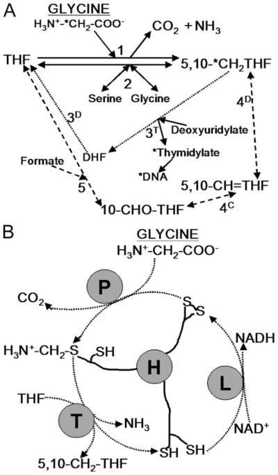
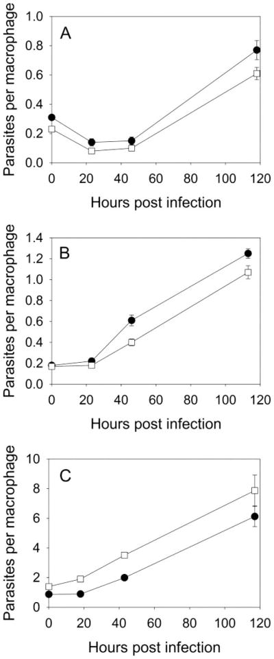
For the human pathogen Leishmania major, a key metabolic function is the synthesis of thymidylate, which requires 5,10-methylenetetrahydrofolate (5,10-CH(2)-THF). 5,10-CH(2)-THF can be synthesized from glycine by the mitochondrial glycine cleavage complex (GCC). Bioinformatic analysis revealed the four subunits of the GCC in the L. major genome, and the role of the GCC in parasite metabolism and virulence was assessed through studies of the P subunit (glycine decarboxylase (GCVP)). First, a tagged GCVP protein was expressed and localized to the parasite mitochondrion. Second, a gcvP(-) mutant was generated and shown to lack significant GCC activity using an indirect in vivo assay after incorporation of label from [2-(14)C]glycine into DNA. The gcvP(-) mutant grew poorly in the presence of excess glycine or minimal serine; these studies also established that L. major promastigotes require serine for optimal growth. Although gcvP(-) promastigotes and amastigotes showed normal virulence in macrophage infections in vitro, both forms of the parasite showed substantially delayed replication and lesion pathology in infections of both genetically susceptible or resistant mice. These data suggest that, as the physiology of the infection site changes during the course of infection, so do the metabolic constraints on parasite replication. This conclusion has great significance to the interpretation of metabolic requirements for virulence. Last, these studies call attention in trypanosomatid protozoa to the key metabolic intermediate 5,10-CH(2)-THF, situated at the junction of serine, glycine, and thymidylate metabolism. Notably, genome-based predictions suggest the related parasite Trypanosoma brucei is totally dependent on the GCC for 5,10-CH(2)-THF synthesis.


Figures

FIGURE 1
FIGURE 1
A, 5,10- CH2-THF formation by the glycine cleavage complex and related folate metabolism in Leishmania. Solid lines indicate direct interconversion of THF and 5,10-CH2-THF (and serine and glycine). The GCC (reaction 1) converts glycine and THF to 5,10-CH2-THF, NH3, and CO2 (NAD+ is also converted to NADH). 5,10-CH2-THF is also formed by SHMT (reaction 2), which is reversible. Dotted and dashed lines indicate indirect routes of THF to 5,10-CH2-THF conversion via dihydrofolate (DHF) and the activity of bifunctional DHFR-TS (reactions 3D and 3T, dotted line) or via 10-formyl-tetrahydrofolate (10-CHO-THF) and 5,10-methenyl-tetrahydrofolate (5,10-CH=THF) using the activity of reversible bifunctional methlenetetrahydrofolate dehydrogenase (4D)-5,10-methenyl-tetrahydrofolate cyclohydrolase (4C) and the activity of 10-formyl-tetrahydrofolate synthase (5) (dashed line). The pathway for incorporation of radiolabel from the 2 position of glycine into DNA is marked by asterisks. B, diagram of glycine cleavage complex. P protein decarboxylates glycine and transfers the amino-methylene moiety to lipoate arm of H protein. T protein releases ammonia and transfers the methylene group to THF, forming 5,10-CH2-THF. The H protein is re-oxidized by lipoate dehydrogenase (L) with concurrent production of NADH.
FIGURE 2
FIGURE 2. GCVP gene disruption
A, gene disruption scheme. Line 1 shows the GCVP gene (open box) in its chromosomal context. Lines 2 and 3 show GCVP disruption constructs. Disruption constructs consisted of the complete GCVP gene interrupted at a PmlI site 734 nucleotides from the 5′ end by autonomous drug resistance cassettes (PAC or HYG, 1.2 or 1.5 kb) consisting of the PAC or HYG open reading frame (gray boxes) whose expression was driven by a segment of 5′-flanking DNA bearing a functional splice acceptor (black boxes). Disruption cassettes were excised from their parental plasmids (pBS-GCVP-PAC and pBS-GCVP-HYG) and introduced into parasites successively. The locations of PCR primers used to confirm the homologous replacement of a WT allele with the PAC or HYG “disruptant” alleles are shown (arrowheads) as is the location of AccI sites and the hybridization probe used in the Southern blot shown in panel B. Dotted lines indicate areas of potential homologous recombination. B, Southern blot of GCVP disruption. DNA from parasite lines digested with AccI was blotted and hybridized with a probe matching the region of DNA indicated in panel A. Lane +/+, WT DNA; lane +/−, WT/gcvP::PAC heterozygote DNA; lane/−, gcvP::PAC/gcvP::HYG double disruption (gcvP). Positions of standards (in kb) and WT and HYG- and PAC-disrupted bands are shown.
FIGURE 3
FIGURE 3. Localization of GCVP-GFP fusion protein
L. major WT-[pXG-GCVP-GFP] promastigotes expressing GCVP-GFP from the pXG-GCVP-GFP plasmid counter-stained with Hoechst 33342. Left panel, GFP fluorescence. Center panel, Hoechst fluorescence. Right panel, GFP and Hoechst fluorescence combined. All fluorescence images are shown overlaid on the phase-contrast image of the parasites.
FIGURE 4
FIGURE 4. Lack of GCVP delays lesion development in mouse infections
Lesion development after inoculation of BALB/c (A and C) or C57/BL6 (B) mouse footpads with 1 × 10⁵ metacyclic (peanut agglutinin minus) promastigotes (A and B) or 1 × 10⁴ amastigotes (C). ●, WT; □, gcvP; ▲, gcvP/+GCVP parasites. Error bars indicate S.E. of mean of measurements of four infected mice. Similar results were seen in five other experiments using BALB/c mice (three with metacyclic parasites and two with amastigotes).
FIGURE 5
FIGURE 5. Macrophage infections with gcvP metacyclic promastigotes or amastigotes
Infection of macrophages in vitro with metacyclic promastigotes (A, high serine medium; B, low serine medium) or amastigotes (C, high-serine medium). ●, WT; □, gcvP. Error bars indicate S.E. from triplicate macrophage coverslip cultures.

References

    1. Herwaldt BL. Lancet. 1999;354:1191–1199. - PubMed
    1. Lira R, Sundar S, Makharia A, Kenney R, Gam A, Saraiva E, Sacks D. J Infect Dis. 1999;180:564–567. - PubMed
    1. Berman J, Bryceson AD, Croft S, Engel J, Gutteridge W, Karbwang J, Sindermann H, Soto J, Sundar S, Urbina JA. Trans R Soc Trop Med Hyg. 2006;100(Suppl 1):41–44. - PubMed
    1. Garrett CE, Coderre JA, Meek TD, Garvey EP, Claman DM, Beverley SM, Santi DV. Mol Biochem Parasitol. 1984;11:257–265. - PubMed
    1. Stechmann A, Cavalier-Smith T. Science. 2002;297:89–91. - PubMed

Publication types

MeSH terms

Substances

LinkOut - more resources