Review
. 2008 Feb;143(2):145-53.
doi: 10.1093/jb/mvm214. Epub 2007 Nov 2.

Packaging the genome: the structure of mitotic chromosomes

Affiliations
Review

Packaging the genome: the structure of mitotic chromosomes

Kazuhiro Maeshima et al. J Biochem. 2008 Feb.

Abstract

Mitotic chromosomes are essential structures for the faithful transmission of duplicated genomic DNA into two daughter cells during cell division. Although more than 100 years have passed since chromosomes were first observed, it remains unclear how a long string of genomic DNA is packaged into compact mitotic chromosomes. Although the classical view is that human chromosomes consist of radial 30 nm chromatin loops that are somehow tethered centrally by scaffold proteins, called condensins, cryo-electron microscopy observation of frozen hydrated native chromosomes reveals a homogeneous, grainy texture and neither higher-order nor periodic structures including 30 nm chromatin fibres were observed. As a compromise to fill this huge gap, we propose a model in which the radial chromatin loop structures in the classic view are folded irregularly toward the chromosome centre with the increase in intracellular cations during mitosis. Consequently, compact native chromosomes are made up primarily of irregular chromatin networks cross-linked by self-assembled condensins forming the chromosome scaffold.


Figures

Fig. 1.
Fig. 1.
(A) A long DNA molecule with a diameter of 2 nm is wrapped around a core histone octamer that consists of H2A, H2B, H3 and H4 histone proteins, and forms a ‘nucleosome’ with a diameter of 11 nm. The nucleosome is assumed to be folded into 30 nm chromatin fibres (1), although the existence of such continuous 30 nm chromatin fibres in native chromosomes remains controversial. Furthermore, it remains unclear how this 30 nm fibre is compacted into the chromatid with a diameter of about 0.7 µm. (B) Schematic representation of a histone-depleted chromosome. The histones were removed from the isolated mitotic chromosome gently by competition with an excess of the polyanions dextran sulfate and heparin. During this process, the positively charged histones in the chromatins are exchanged for the highly negatively charged polyanions. After removing the histones, the DNA remains highly organized by the ‘chromosome scaffold’ (drawn in red), keeping the size and shape of the original chromosomes (9). (C) ‘Hierarchical helical folding model’. This model assumes that the 30 nm chromatin fibres are folded into 100 nm fibres and then progressively into 200–250 nm fibres that coil to form the final mitotic chromosomes. (D) Major chromosome scaffold components have axial distributions at the centere of each chromatid in the compact chromosomes. An isolated human chromosome was stained using antibodies for topoisomerase IIα and the condensin I component hCAP-H. The antibody labelling shows axial structures with a diameter of ∼200 nm within the chromosome body stained with DAPI. Note that the staining for topoisomerase IIα and condensin I is localized in an alternating manner, forming a ‘barber pole’ structure (26). The images are adapted from (26) with the permission of Elsevier.
Fig. 2.
Fig. 2.
(A) and (B) Conventional electron microscopy views of isolated chromosomes, swollen in a low-salt buffer containing 1 mM Mg2+. Uniform 30 nm chromatin fibre loops that diverge radially from the centre are clearly visualized in cross- (A) and longitudinal- (B) sections of the chromosomes (19). The chromosomes seem to consist of radial chromatin loops that are somehow tethered centrally by the scaffolding protein condensin I, mapped using immuno-gold (shown by arrows). The images are reproduced from (19) with the permission of Springer. The bar indicates 200 nm. (C) Cryo-EM images of a frozen-hydrated section of mitotic HeLa cells. The compact areas, outlined by the dashed line, are cross-sections of mitotic chromosomes (Xs), which are surrounded by cytoplasm full of electron-dense ribosomes and other particles. The selected area is magnified in (D). Note the grainy, homogeneous texture of the chromosome (Xs). No higher-order or periodic structures are recognized, such as 30 nm chromatin fibres. The scale bar indicates 500 nm in (C) and 100 nm in (D).
Fig. 3.
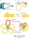
Fig. 3.
(A) Schematic representation of the structure of condensin. In vertebrates, there are two types of condensin, condensin I and condensin II. Condensin I consists of five different subunits, a heterodimer of SMC4 (CAP-C) and SMC2 (CAP-E) and three non-SMC subunits: CAP-D2 (Eg-7), and CAP-G, and CAP-H (Kleisin γ, Barren). In vertebrates, condensin II has the SMC2 and SMC4 heterodimer in common and three distinct non-SMC subunits: CAP-D3, CAP-G2, and CAP-H2 (Kleisin γ). (B) Condensins can introduce positive supercoils into closed circular DNA via ATP hydrolysis. However, it is not known how this condensin activity functions in the condensation process. (C) A proposed model of the mitotic chromosome structure. A cross-section of an isolated chromosome swollen in a low-salt buffer shows radial chromatin loops that are somehow tethered centrally by condensin (Figs 2A and B). Our model supposes two events for building a robust chromosomal architecture. First, condensins bind to certain specific sites in the genome chromatin to make loops (loop-forming activity). Second, condensins further cross-link neighbouring chromatin loops (networking activity) and form anisotropic self-assembly structures in a cooperative manner, like a scaffold. With the increase in intracellular cations during mitosis, the loop structures fold irregularly toward the chromosome scaffold containing abundant condensins. Due to this collapsing-loop process, compact native chromosomes are made up primarily of irregular chromatin networks cross-linked by self-assembled condensins, forming the chromosome scaffold. Note that no continuous 30 nm chromatin fibres are visible in the compact native chromosomes. In the absence of condensins (49), chromosomes are still condensed by RCA, but with loss of their structural integrity.

References

    1. Kornberg RD, Lorch Y. Twenty-five years of the nucleosome, fundamental particle of the eukaryote chromosome. Cell. 1999;98:285–294. - PubMed
    1. Luger K, Mader AW, Richmond RK, Sargent DF, Richmond TJ. Crystal structure of the nucleosome core particle at 2.8 A resolution. Nature. 1997;389:251–260. - PubMed
    1. Robinson PJ, Fairall L, Huynh VA, Rhodes D. EM measurements define the dimensions of the “30-nm” chromatin fiber: evidence for a compact, interdigitated structure. Proc. Natl. Acad. Sci. USA. 2006;103:6506–6511. - PMC - PubMed
    1. Finch JT, Klug A. Solenoidal model for superstructure in chromatin. Proc. Natl. Acad. Sci. USA. 1976;73:1897–1901. - PMC - PubMed
    1. Bednar J, Horowitz RA, Dubochet J, Woodcock CL. Chromatin conformation and salt-induced compaction: three-dimensional structural information from cryoelectron microscopy. J. Cell Biol. 1995;131:1365–1376. - PMC - PubMed

Publication types