. 2007;35(22):7636-50.
doi: 10.1093/nar/gkm931. Epub 2007 Nov 2.

Regulatory mechanisms for 3'-end alternative splicing and polyadenylation of the Glial Fibrillary Acidic Protein, GFAP, transcript

Affiliations

Regulatory mechanisms for 3'-end alternative splicing and polyadenylation of the Glial Fibrillary Acidic Protein, GFAP, transcript

Jenny Blechingberg et al. Nucleic Acids Res. 2007.

Abstract

The glial fibrillary acidic protein, GFAP, forms the intermediate cytoskeleton in cells of the glial lineage. Besides the common GFAP alpha transcript, the GFAP epsilon and GFAP kappa transcripts are generated by alternative mRNA 3'-end processing. Here we use a GFAP minigene to characterize molecular mechanisms participating in alternative GFAP expression. Usage of a polyadenylation signal within the alternatively spliced exon 7a is essential to generate the GFAP kappa and GFAP kappa transcripts. The GFAP kappa mRNA is distinct from GFAP epsilon mRNA given that it also includes intron 7a. Polyadenylation at the exon 7a site is stimulated by the upstream splice site. Moreover, exon 7a splice enhancer motifs supported both exon 7a splicing and polyadenylation. SR proteins increased the usage of the exon 7a polyadenylation signal but not the exon 7a splicing, whereas the polypyrimidine tract binding (PTB) protein enhanced both exon 7a polyadenylation and exon 7a splicing. Finally, increasing transcription by the VP16 trans-activator did not affect the frequency of use of the exon 7a polyadenylation signal whereas the exon 7a splicing frequency was decreased. Our data suggest a model with the selection of the exon 7a polyadenylation site being the essential and primary event for regulating GFAP alternative processing.


Figures
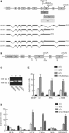
Figure 1.
Figure 1.
Construction and functional analysis of a GFAP minigene. (A) A schematic picture of the GFAP gene structure. The grey boxes denote the exons and the black horizontal lines the introns. Below is shown the arrangement of the exons in the GFAPα, GFAPε, GFAPκ, GFAPδ and GFAPΦ mRNA. The positions of stop codons are indicated by asterisks. (B) A schematic picture of the GFAP wt minigene. GFAP gene sequences from intron 6 through exon 9 were inserted into the pTAG4 vector. The vector contains the SV40 promoter and two internal exons (marked 1 and 2) and lacks internal poly(A) signals. The positions of the reverse primers used for real time PCR are denoted as black arrows: primers in intron 7a, exon 7a, intron 7 and exon 9 for amplification of GFAPκ, GFAPε, GFAPδ and GFAPΦ, and GFAPα, respectively. The forward PCR primer was spanning exon 1 and 2. (C) Gel electrophoresis of the PCR products from real-time PCR with the primers shown in (B). GFAPα (266 bp), GFAPε (299 bp) and GFAPκ (296 bp) are expressed from the GFAP wt minigene. Since the GFAPκ mRNA is 354 bp longer than the GFAPε mRNA, only GFAPε is amplified by the primer placed in exon 7a, at the used experimental conditions. (D) The expressed ratios of GFAPα/GFAPε, GFAPα/GFAPκ and GFAPε/GFAPκ mRNA in normal human astrocytes (NHA cells), primary rat astrocytes (PRA), A172, HeLa, N2A and HEK293T cells transiently transfected with the GFAP wt minigene. The SDs from three independent assays are shown. The E7pA/E9pA ratios are also shown. (E) The expressed ratios of GFAPα/GFAPε, GFAPα/GFAPκ and GFAPε/GFAPκ mRNA, as well as the E7pA/E9pA ratios, in A172 cells transiently transfected with the GFAP wt, the GFAP+ATG (coding) or the GFAP+1828 bp minigenes. The E7pA/E9pA ratios are also shown.
Figure 2.
Figure 2.
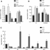
Determination of the relative GFAP mRNA stabilities. N2A cells transfected with the GFAP minigene were 48 h after transfection added actinomycin-D or as control the vehicle solution DMSO for the indicated time points. RNA was extracted and real-time RT–PCR was used to determine the ratios of GFAPα/GFAPε mRNA, GFAPα/GFAPκ mRNA and GFAPε/GFAPκ mRNA, as well as the E7pA/E9pA ratio. The labelling H2O indicates transfected N2A cells not treated with DMSO and actinomycin-D.
Figure 3.
Figure 3.
Environmental conditions affect GFAP polyadenylation signal selection. (A) N2A cells transfected with the GFAP minigene were added BAPTA or as control the vehicle solution NaHCO3. RNA was extracted and real-time RT–PCR was used to determine the ratios of GFAPα/GFAPε mRNA, GFAPα/GFAPκ mRNA and GFAPε/GFAPκ mRNA, as well as the E7pA/E9pA ratio. (B) As in (A) except for the addition of CdCl2 or as control H2O. (C) Transfected N2A cells were 24 h after transfection incubated at 42°C for the indicated time points. RNA was analysed by real-time RT–PCR as described in (A).
Figure 4.
Figure 4.
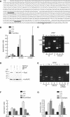
Exon 7a polyadenylation is required for alternative GFAP 3′-end processing. (A) The sequence of the polyadenylation signal in exon 7a with the AAUAAA hexamer and the AUUAAA hexamer (underlined). The position of poly(A) tail additions with origin from the polyadenylation signals were identified by 3′-RACE experiments and are marked with underscore. The G-rich region downstream the poly(A) signal compositing the PRE motif is shown in italic and bold. This region together with flanking sequences was deleted in the dPRE minigene. The position of the examined SNP is double underlined. The position of the intron 7a/exon 7a border is marked by a slash. (B) A172 cells were transiently transfected with the GFAP wt, GFAP-mE7pA (mutation of the exon 7a polyadenylation signal) or the GFAP-mE9pA (mutation of the exon 9 polyadenylation signal) minigenes. The expressed ratios were quantified by real-time PCR. (C) Gel electrophoresis of RT–PCR performed with cDNA made of total RNA extracted from A172 cells transiently transfected with either the GFAP wt minigene (wt) or GFAP-mE7pA. GFAPα (266 bp) and GFAPε (299 bp) are detected in (wt) and GFAP-mE7pA transfected cells. Note that the primers for GFAPε in this experimental setting also amplify cDNA representing GFAPκ (653 bp) (indicated by arrow). Neither GFAPδ nor GFAPΦ expression could be detected (expected sizes of 951 and 1305 bp, respectively). Plasmid DNA is used as a positive control for the reverse primer used for GFAPΦ amplification (lane C). The position of the reverse primers is schematically shown in Figure 1B. (D) Western blot of protein extracted from HEK293T cells transfected with either the GFAP-mE7pA minigene alone, or together with siRNAs against Rrp6 (siRrp6) or an unspecific siRNA (SiC) as control. To the left the marker sizes are shown. Expression of Rrp6 (100 kDa) is detected with a monoclonal antibody and as loading control the expression of hnRNPC1/C2 is shown (33 kDa). (E) RT–PCR of RNA extracted from the transfected HEK293T cells in (D). GFAPα and GFAPε and are detected as fragments of 266 and 299 bp, respectively. Neither GFAPδ nor GFAPΦ are detected (expected sizes of 951 bp and 1305 bp, respectively), in wt, GFAP-mE7pA, or GFAP-mE7pA and siRrp6 transfected cells. Plasmid DNA is used as a positive control for the reverse primer used for GFAPΦ amplification (lane C). The position of the reverse primers is schematically shown in Figure 1B. (F) Cis-elements influence alternative GFAP polyadenylation signal selection. Primary rat astrocytic cells were transiently transfected with either GFAP wt or GFAP-dPRE (deleted PRE sequence) minigenes. The expression ratios were quantified by real-time RT–PCR as previously described. (G) A minigene with nucleotide A in the SNP position 6 in the PRE motif was examined together with the wt minigene as in (F). Note the wt minigene contains a G nucleotide in the PRE SNP.
Figure 5.
Figure 5.
Exon 7a polyadenylation requires a functional 5′-splice site. (A) An overview of the pyrimidine tract preceding the 5′-splice site of exon 7a. The branch point consensus is shown to the left, and the consensus sequence of the ‘ideal’ pyrimidine tract is shown beneath the exon 7a sequence. Below, the sequence of the mutated, optimized sequence is shown. (B) The expressed ratios of GFAPα/GFAPε, GFAPα/GFAPκ and GFAPε/GFAPκ, together with the E7pA/E9pA ratio, in N2A cells transiently transfected with the GFAP wt, GFAP-d3'ss gene with a mutated intron 7a 3′-splice site, or the GFAP-Y minigene with an optimized polypyrimidine tract in the intron 7a 3′-splice site.
Figure 6.
Figure 6.
Exon 7a splicing and polyadenylation is regulated by SR proteins and exon splice enhancer, ESE, sequences. (A) The sequence of the 5′-part of exon 7a. The three putative ESEs are underlined. In order to test their functionality they were mutated either individually or in combination to produce the minigenes ESE1, ESE2, ESE3 and ESE1/3, respectively. (B) The expressed ratios of GFAPα/GFAPε, GFAPα/GFAPκ and GFAPε/GFAPκ, and the E7pA/E9pA ratio, in N2A cells transiently transfected with the GFAP wt or the ESE1, ESE2, ESE3 and ESE1/3 minigenes. (C) As (B) but with transfections in A172 cells. (D) The expressed ratios in N2A cells transiently transfected with the GFAP wt, GFAP-Y or the GFAP-Y-ESE1/3 combined mutation.
Figure 7.
Figure 7.
Trans-factors influence alternative GFAP polyadenylation signal selection. (A) Co-expression of the GFAP wt minigene and trans-factors involved in polyadenylation signal selection. A172 cells were transiently transfected with either the GFAP wt minigene and an empty vector or the GFAP wt minigene and a vector containing the open reading frame of factors determined to be involved in alternative polyadenylation. The GFAP mRNA ratios were quantified by real-time PCR. C indicates the control experiment in where the GFAPwt minigene was cotransfected with empty expression vector. (B) The GFAP mRNA ratios were determined in A172 cells transiently transfected with GFAP wt together with plasmids encoding the SR proteins ASF/SF2, SC35, Srp40, 9G8 or Srp55. C indicates the control experiment in where the GFAPwt minigene was cotransfected with empty expression vector. As a control RNA binding protein was included a transfection with an hnRNPF expression vector which in (A) was shown to have no effect on the expression ratios. The expressed ratios were quantified by real-time PCR.
Figure 8.
Figure 8.
Promoter activity influences alternative GFAP 3′-end processing. (A and B) The SV40 promoter in the GFAP wt minigene was switched with the endogenous GFAP promoter and transiently transfected into NHA cells (A) or primary rat astrocytes (B). The indicated expressed ratios were quantified by real-time PCR. (C) The SV40 promoter in the GFAP wt minigene was switched with the β-globin minimal promoter or the tk minimal promoter with additional two upstream GAL4 recognition sites. The minigenes were transiently transfected into N2A cells co-transfected with the chimerical transcription factor GAL4-VP16. The expressed ratios were quantified by real-time PCR. Note that for illustration purposes the GFAPκ/GFAPε ratio is shown in this particular figure.

References

    1. Eng LF, Ghirnikar RS, Lee YL. Glial fibrillary acidic protein: GFAP-thirty-one years (1969–2000) Neurochem Res. 2000;25:1439–1451. - PubMed
    1. Geisler N, Weber K. The amino acid sequence of chicken muscle desmin provides a common structural model for intermediate filament proteins. EMBO J. 1982;1:1649–1656. - PMC - PubMed
    1. Parry DA, Steinert PM. Intermediate filament structure. Curr. Opin. Cell. Biol. 1992;4:94–98. - PubMed
    1. Zelenika D, Grima B, Brenner M, Pessac B. A novel glial fibrillary acidic protein mRNA lacking exon 1. Brain Res. Mol. Brain Res. 1995;30:251–258. - PubMed
    1. Condorelli DF, Nicoletti VG, Barresi V, Conticello SG, Caruso A, Tendi EA, Giuffrida Stella AM. Structural features of the rat GFAP gene and identification of a novel alternative transcript. J. Neurosci. Res. 1999;56:219–228. - PubMed

Publication types

MeSH terms