

Is arginine charged in a membrane?
- PMID: 17981901
- PMCID: PMC2157258
- DOI: 10.1529/biophysj.107.121566
Is arginine charged in a membrane?
Abstract
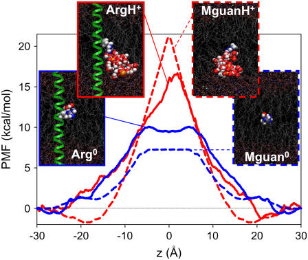
"Charged" amino acids play countless important roles in protein structure and function. Yet when these side chains come into contact with membranes we do not fully understand their behavior. This is highlighted by a recent model of voltage-gated ion channel activity and translocon-based experiments that suggest small penalties to expose these side chains to lipids, opposing the prevailing view in membrane biophysics. Here we employ a side chain analog as well as a transmembrane helix model to determine the free energy as a function of protonation state and position for a lipid-exposed arginine (Arg) residue across a membrane. We observe high free energy barriers for both the charged and neutral states. Due to the stabilizing influence of membrane deformations for the protonated form, the Arg side chain experiences a pK(a) shift of <or=4.5 units and remains mostly protonated. The cost for exposing Arg to lipid hydrocarbon is prohibitively high with implications for many membrane translocating processes and the activation mechanisms of voltage-gated ion channels.
Figures

References
-
- Nozaki, Y., and C. Tanford. 1967. Examination of titration behavior. Methods Enzymol. 11:715–734.
-
- Angyal, S. J., and W. K. Warburton. 1951. The basic strengths of methylated guanidines. J. Chem. Soc. 1951:2492–2494.
-
- Hall, N. F., and M. R. Sprinkle. 1932. Relations between the structure and strength of certain organic bases in aqueous solution. J. Am. Chem. Soc. 54:3469–3485.
-
- Brown, K. L., and R. E. Hancock. 2006. Cationic host defense (antimicrobial) peptides. Curr. Opin. Immunol. 18:24–30. - PubMed
-
- Kalderon, D., W. D. Richardson, A. F. Markham, and A. E. Smith. 1984. Sequence requirements for nuclear localisation of SV40 large-T antigen. Nature. 311:33–38. - PubMed
Publication types
MeSH terms
Substances
Grants and funding
LinkOut - more resources
Full Text Sources
