. 2008 Jan;146(1):213-27.
doi: 10.1104/pp.107.105890. Epub 2007 Nov 2.

Glufosinate ammonium-induced pathogen inhibition and defense responses culminate in disease protection in bar-transgenic rice

Affiliations

Glufosinate ammonium-induced pathogen inhibition and defense responses culminate in disease protection in bar-transgenic rice

Il-Pyung Ahn. Plant Physiol. 2008 Jan.

Abstract

Glufosinate ammonium diminished developments of rice (Oryza sativa) blast and brown leaf spot in 35S:bar-transgenic rice. Pre- and postinoculation treatments of this herbicide reduced disease development. Glufosinate ammonium specifically impeded appressorium formation of the pathogens Magnaporthe grisea and Cochliobolus miyabeanus on hydrophobic surface and on transgenic rice. In contrast, conidial germination remained unaffected. Glufosinate ammonium diminished mycelial growth of two pathogens; however, this inhibitory effect was attenuated in malnutrition conditions. Glufosinate ammonium caused slight chlorosis and diminished chlorophyll content; however, these alterations were almost completely restored in transgenic rice within 7 d. Glufosinate ammonium triggered transcriptions of PATHOGENESIS-RELATED (PR) genes and hydrogen peroxide accumulation in transgenic rice and PR1 transcription in Arabidopsis (Arabidopsis thaliana) wild-type ecotype Columbia harboring 35S:bar construct. All transgenic Arabidopsis showed robust hydrogen peroxide accumulation by glufosinate ammonium. This herbicide also induced PR1 transcription in etr1 and jar1 expressing bar; however, no expression was observed in NahG and npr1. Fungal infection did not alter transcriptions of PR genes and hydrogen peroxide accumulation induced by glufosinate ammonium. Infiltration of glufosinate ammonium did not affect appressorium formation of M. grisea in vivo but inhibited blast disease development. Hydrogen peroxide scavengers nullified blast protection and transcriptions of PR genes by glufosinate ammonium; however, they did not affect brown leaf spot progression. In sum, both direct inhibition of pathogen infection and activation of defense systems were responsible for disease protection in bar-transgenic rice.


Figures
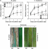
Figure 1.
Figure 1.
Effects of glufosinate ammonium (G) on the developments of rice blast and brown leaf spot caused by M. grisea strain KJ201 and C. miyabeanus strain HIH-1, respectively. Glufosinate ammonium or mock was applied 24 h before fungal inoculation. Disease progression was estimated according to the number and size of lesions. A, Disease progression in ‘Dongjin’ (−, NC) and bar-transgenic ‘Dongjin’ (+) pretreated with mock (−, 250 μg mL−1 Tween 20) or glufosinate ammonium (+, 100 μg mL−1) and inoculated with M. grisea strain KJ201 and C. miyabeanus strain HIH-1. Each data point is mean ± se. B, Blast and brown leaf spot developments in ‘Dongjin’ (NC) and bar-transgenic ‘Dongjin’ (bar) pretreated with glufosinate ammonium (+) or mock (−). Photographs depicting representative symptoms were taken 10 d after fungal inoculation. Experiments were repeated more than three times with three replicates consisting of 15 plants; almost similar tendencies were obtained. [See online article for color version of this figure.]
Figure 2.
Figure 2.
Rice blast and rice brown leaf spot disease progressions of glufosinate ammonium-treated bar-transgenic (bar) or NC rice plants. Plants were inoculated by M. grisea strain KJ201 or C. miyabeanus strain HIH-1. In the meantime, 100 μg mL−1 of glufosinate ammonium was sprayed 5 d or 1 d prior to (preinoculation treatments; −5 and −1, respectively) fungal inoculation. Treatments were also performed 12 h or 1 d after (postinoculation treatments; +0.5 and +1, respectively) inoculation. In the analysis of preinoculation treatment effects, glufosinate ammonium-treated rice leaves were carefully washed several times by spraying distilled water at 24 h after treatment. Disease progression was determined 7 dpi. Each data point is mean ± se. Different letters indicate statistically significant differences between treatments (Duncan's multiple range test; P < 0.05). Experiments were repeated more than three times with three replicates consisting of 15 rice plants; almost similar tendencies were observed.
Figure 3.
Figure 3.
Effects of glufosinate ammonium on conidial germination and appressorium formation in M. grisea strain KJ201 and C. miyabeanus strain HIH-1. Conidial germination and appressorium formation were estimated microscopically 24 h after placement onto the tested surface. A, Effects of glufosinate ammonium on prepenetration development of M. grisea and C. miyabeanus on the hydrophobic surface of GelBond. Each data point is mean ± se. B, Effects of glufosinate ammonium (100 μg mL−1) on conidial germination (white bar) and appressorium formation (black bar) of M. grisea and C. miyabeanus on rice leaves from bar-transgenic plants. Different letters indicate statistically significant differences between treatments (Duncan's multiple range test; P < 0.05). C, Prepenetration developments of M. grisea and C. miyabeanus on the hydrophobic surface of GelBond (artificial surface) or transgenic (bar) rice leaves in the presence (+) or absence (−) of 100 μg mL−1 glufosinate ammonium. a, Appressorium; c, conidium; g, germ tube. Bars = 50 μm. Experiments were repeated more than three times with three replicates; almost similar tendencies were obtained. [See online article for color version of this figure.]
Figure 4.
Figure 4.
Effects of nutrient starvation and/or glufosinate ammonium on the fungal growth. Prior to colony contact on the CM, colony morphologies of M. grisea (A) and C. miyabeanus (B) on the CM containing carbon and nitrogen sources (+) or mock (−) and supplemented with glufosinate (+) or mock (−) were photographed and colony areas were measured (C). Different letters indicate statistically significant differences between treatments (Duncan's multiple range test; P < 0.05). Experiments were repeated more than three times with three replicates; almost similar results were obtained. [See online article for color version of this figure.]
Figure 5.
Figure 5.
Transcriptions of PR genes induced by glufosinate ammonium (100 μg mL−1) treatment and/or pathogen inoculation at varying dpt and dpi. Pathogen inoculation and glufosinate ammonium treatment were performed as described in Figure 1A. A, PR1 gene transcription induced by varying concentrations of glufosinate ammonium treatment at 1 dpt. B, Transcription of PR1 gene induced by glufosinate ammonium in bar-transgenic rice. Glufosinate ammonium was sprayed on second leaves. Leaves sprayed with glufosinate ammonium (L, local) or left untreated (S, systemic) were harvested from the same plants 1 dpt. Transgenic rice leaves treated with mock (C) were also harvested at the same time. C, PR1 gene expression was retained up to 15 d after glufosinate treatment. D, Transcriptions of PR1, PBZ1, and POX22.3 in NC and transgenic (bar) rice challenged with M. grisea strain KJ201 or C. miyabeanus strain HIH-1 1 d after mock or 100 μg mL−1 glufosinate ammonium. Total RNA was extracted from the leaves of five rice plants recovered 0, 1, 2, 3, and 4 dpi. Experiments were repeated more than three times; almost similar results were obtained.
Figure 6.
Figure 6.
Effects of glufosinate ammonium on the accumulation of hydrogen peroxide and transcriptions of PR1 and PDF1.2 in 35S:bar-transgenic Arabidopsis Col-0 and its mutants. Glufosinate (+, 100 μg mL−1) was sprayed onto rosette leaves and treated rosette and nontreated cauline leaves were harvested 24 h later. A, Hydrogen peroxide accumulation in treated rosette (local) and nontreated cauline (systemic) leaves of 35S:bar-harboring transgenic Arabidopsis Col-0, NahG, npr1, etr1, and jar1 lines exposed to glufosinate. Harvested leaves were stained with DAB (0.1%, w/v). B, Quantification of hydrogen peroxide accumulation in treated rosette (local) and nontreated (systemic) cauline leaves of transgenic Arabidopsis Col-0, NahG, npr1, etr1, and jar1 lines exposed to glufosinate. Each bar represents the mean ± se. Different letters indicate statistically significant differences between treatments (Duncan's multiple range test; P < 0.05). C, Transcriptions of PR1 and PDF1.2 in treated rosette (L; local) and nontreated cauline (S; systemic) leaves of transgenic Arabidopsis Col-0, NahG, npr1, etr1, and jar1. Data were from Arabidopsis sprayed with glufosinate (+) or 250 μg mL−1 Tween 20 only (−, mock). In addition, leaves of transgenic Col-0 were harvested 1 d after salicylic acid (SA, 500 μm) or jasmonic acid (JA, 100 μm) spray. D, Expression of eGFP by glufosinate treatment in transgenic Arabidopsis containing PR1:eGFP and 35S:bar gene construct. Bar = 1 mm. E, Transcription of eGFP transcript under the control of PR1 promoter by glufosinate treatment in transgenic Arabidopsis. Experiments were repeated more than three times; almost similar results were obtained.
Figure 7.
Figure 7.
Effects of glufosinate ammonium and M. grisea strain KJ201 on hydrogen peroxide accumulation and fungal ramification in bar-transgenic and nontransgenic rice. The plants were sprayed with glufosinate ammonium (+) in 250 μg mL−1 Tween 20 or mock only (−). One day after treatment, rice plants were inoculated with virulent M. grisea strain KJ201. A, Effects of glufosinate ammonium on the NC and transgenic (bar) rice plants. The 4-week-old plants were sprayed with 100 μg mL−1 glufosinate ammonium (+) or mock (−). Representative leaves were photographed 10 dpt. B, Effects of glufosinate ammonium on the Fv/Fm in NC and transgenic (bar) rice. Each data point is mean ± se. Effects of glufosinate ammonium on hydrogen peroxide accumulation (C) and fungal ramification (D). Microscopic observation of hydrogen peroxide and in planta growth was performed on leaves recovered at 24 and 72 hpi. *, Hydrogen peroxide accumulation. a, Appressorium; c, conidium; m, invasive mycelia. Bars = 50 μm. Experiments were repeated more than three times; almost similar tendencies were obtained.
Figure 8.
Figure 8.
Effects of ascorbic acid and catalase on rice blast and rice brown leaf spot diseases inhibited by glufosinate ammonium on bar-transgenic rice and transcriptions of PR genes. A, Transgenic rice was sprayed with 100 μg mL−1 glufosinate ammonium (+) in 250 μg mL−1 Tween 20 or mock only (−, 250 μg mL−1 Tween 20) and then mock (−, distilled water), ascorbic acid (+), or catalase (+) was infiltrated into the carefully washed leaves 24 h after herbicide treatment. M. grisea or C. miyabeanus was inoculated 3 h after treatment with hydrogen peroxide scavengers. Photographs were taken 7 dpi. B, Quantification of rice blast (white bar) and rice brown leaf spot (black bar) disease developments and effects of catalase and ascorbic acid on the appressorium formation in M. grisea (white bar) and C. miyabeanus (black bar). Different letters indicate statistically significant differences between treatments (Duncan's multiple range test; P < 0.05). C, Effects of catalase (+) on hydrogen peroxide accumulation in glufosinate ammonium-treated plants. *, Hydrogen peroxide accumulation. a, Appressorium; c, conidium. Bars = 50 μm. D, Invasive mycelial growth in 35S:bar-transgenic rice pretreated with 100 μg mL−1 glufosinate ammonium (+) and infiltrated with 5,000 units mL−1 catalase (+). M. grisea was inoculated 24 h after final treatment. Photograph depicting representative infection hyphae in aniline blue staining 72 hpi. m, Invasive mycelial growth. Bars = 50 μm. E, Analyses of PR1, PBZ1, and POX22.3 transcriptions in 35S:bar-transgenic rice leaves sprayed with glufosinate ammonium (+) and/or infiltrated with catalase (−). Total RNA was prepared from five plants 24 h after infiltration, separated using denaturing gel electrophoresis, and transferred to nylon membrane. The blots were hybridized with radiolabeled PR1, PBZ1, and POX22.3 probes. All experiments were done at least three times; almost similar results were obtained.
Figure 9.
Figure 9.
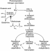
Proposed model for glufosinate-induced disease resistance. Glufosinate initiates accumulation of free radicals by irreversible binding with and inactivation of Gln synthetase. Toxic ammonia derived from photorespiration or nitrogen assimilation is increased within the cell and disturbed electron transport system within chloroplast. Free radicals were produced and in turn, this molecule triggers disease resistance against M. grisea and C. miyabeanus.

References

    1. Agrawal GK, Jwa NS, Iwahashi H, Rakwal R (2003) Importance of ascorbate peroxidases OsAPX1 and OsAPX2 in the rice pathogen response pathways and growth and reproduction revealed by their transcriptional profiling. Gene 322 93–103 - PubMed
    1. Ahn IP, Kim S, Kang S, Suh SC, Lee YH (2005. a) Rice defense mechanisms against Cochliobolus miyabeanus and Magnaporthe grisea are distinct. Phytopathology 95 1248–1255 - PubMed
    1. Ahn IP, Kim S, Lee YH (2005. b) Vitamin B1 functions as an activator of plant disease resistance. Plant Physiol 138 1505–1515 - PMC - PubMed
    1. Ahn IP, Kim S, Lee YH, Suh SC (2007. a) Vitamin B1-induced priming is dependent on hydrogen peroxide and the NPR1 gene in Arabidopsis. Plant Physiol 143 838–848 - PMC - PubMed
    1. Ahn IP, Lee SW, Suh SC (2007. b) Rhizobacteria-induced priming in Arabidopsis is dependent on ethylene, jasmonic acid and NPR1. Mol Plant Microbe Interact 20 759–768 - PubMed

Publication types

MeSH terms