. 2007 Nov;19(11):3723-38.
doi: 10.1105/tpc.107.053371. Epub 2007 Nov 2.

Glycolytic enzymes associate dynamically with mitochondria in response to respiratory demand and support substrate channeling

Affiliations

Glycolytic enzymes associate dynamically with mitochondria in response to respiratory demand and support substrate channeling

James W A Graham et al. Plant Cell. 2007 Nov.

Abstract

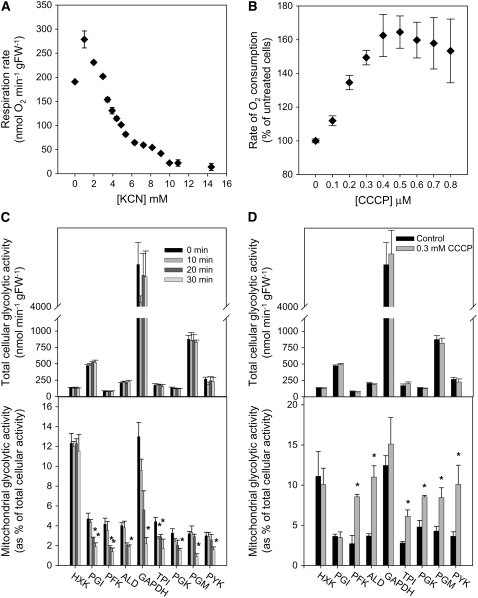
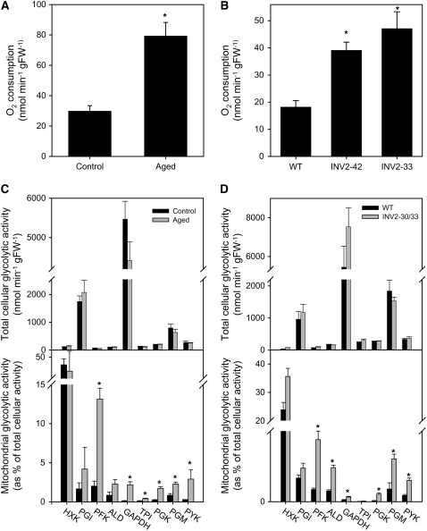
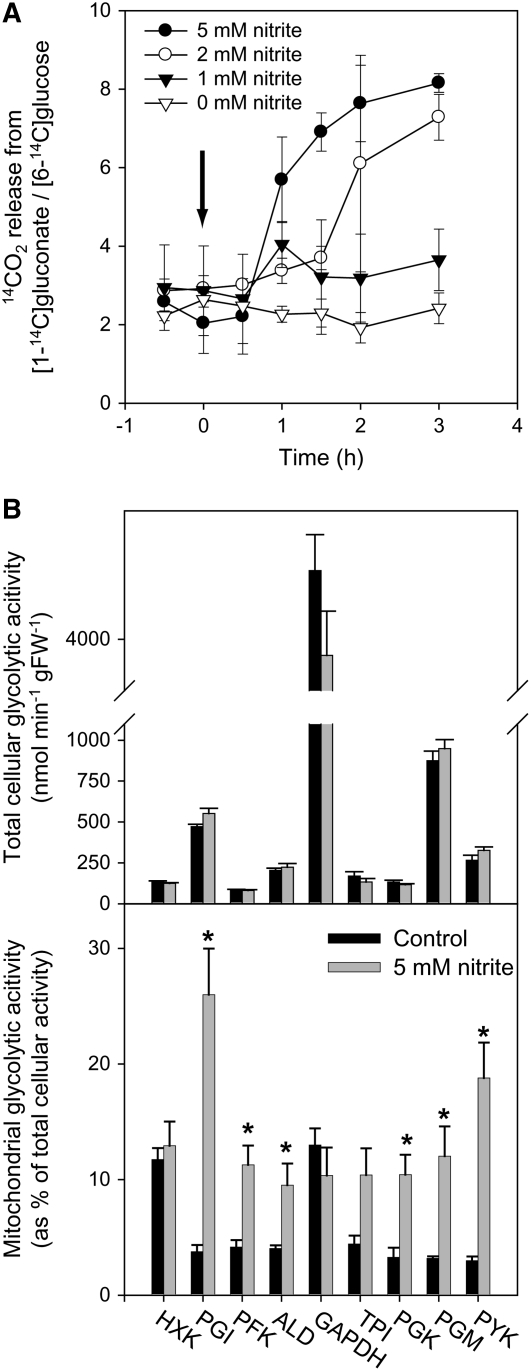
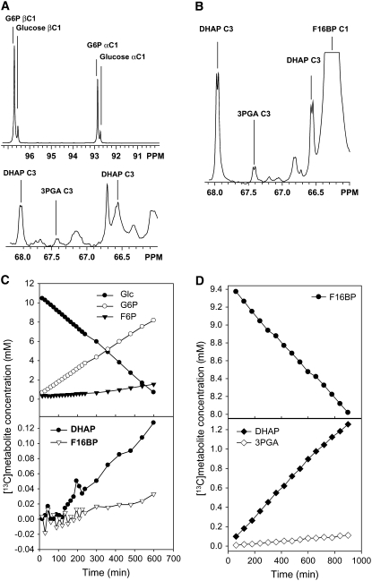
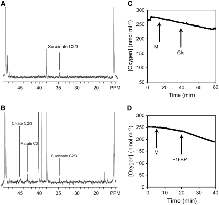
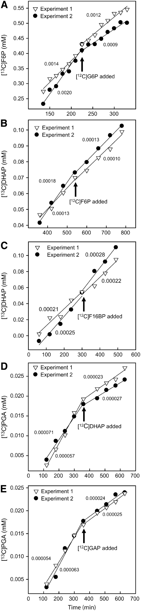
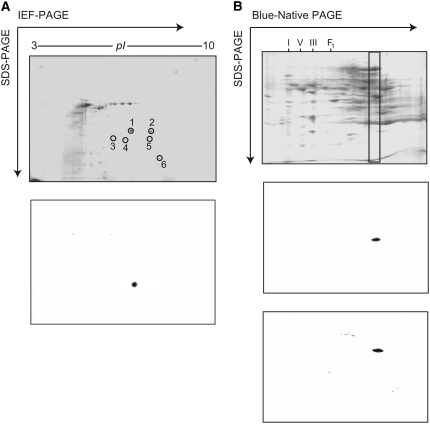
In Arabidopsis thaliana, enzymes of glycolysis are present on the surface of mitochondria and free in the cytosol. The functional significance of this dual localization has now been established by demonstrating that the extent of mitochondrial association is dependent on respiration rate in both Arabidopsis cells and potato (Solanum tuberosum) tubers. Thus, inhibition of respiration with KCN led to a proportional decrease in the degree of association, whereas stimulation of respiration by uncoupling, tissue ageing, or overexpression of invertase led to increased mitochondrial association. In all treatments, the total activity of the glycolytic enzymes in the cell was unaltered, indicating that the existing pools of each enzyme repartitioned between the cytosol and the mitochondria. Isotope dilution experiments on isolated mitochondria, using (13)C nuclear magnetic resonance spectroscopy to monitor the impact of unlabeled glycolytic intermediates on the production of downstream intermediates derived from (13)C-labeled precursors, provided direct evidence for the occurrence of variable levels of substrate channeling. Pull-down experiments suggest that interaction with the outer mitochondrial membrane protein, VDAC, anchors glycolytic enzymes to the mitochondrial surface. It appears that glycolytic enzymes associate dynamically with mitochondria to support respiration and that substrate channeling restricts the use of intermediates by competing metabolic pathways.


Figures

Figure 1.
Figure 1.
Effect of Manipulation of Respiration Rate in Arabidopsis Cell Suspensions on the Degree of Association of Glycolytic Enzymes with Mitochondria. KCN (A) or CCCP (B) was added to heterotrophic Arabidopsis cell suspension cultures at the final concentrations indicated and the rate of oxygen consumption measured until a steady rate was obtained. Arabidopsis cell suspensions were treated with 5 mM KCN for the indicated time interval (C) or 0.3 μM CCCP for 30 min (D). Following these treatments, mitochondria were isolated, and the activities of glycolytic enzymes in the mitochondrial fraction relative to the total cellular activity of each enzyme were determined. HXK, hexokinase; PGI, phosphoglucose isomerase; PFK, phosphofructokinase; ALD, aldolase; GAPDH, glyceraldehyde-3-phosphate dehydrogenase; TPI, triose phosphate isomerase; PGK, phosphoglycerate kinase; PGM, phosphoglyceromutase; PYK, pyruvate kinase; FW, fresh weight. Values are mean of four independent samples ± se. Asterisks indicate significant difference from untreated control (t test; P < 0.05).
Figure 2.
Figure 2.
Effect of Manipulation of Respiration Rate in Potato Tubers on the Degree of Association of Glycolytic Enzymes with Mitochondria. (A) Respiration rate of potato tuber discs after ageing for 16 h. (B) Respiration rate of potato tuber discs from transgenic tubers expressing a yeast invertase in the cytosol. Data shown are pooled from two tubers each of lines INV2-30 and INV2-33 (Roessner et al., 2001). (C) Activities of glycolytic enzymes associated with mitochondria isolated from aged tubers relative to the total cellular activity of each enzyme. (D) Activities of glycolytic enzymes associated with mitochondria isolated from transgenic tubers expressing yeast invertase, relative to the total cellular activity of each enzyme. Abbreviations are as given in Figure 1. Data are the mean of four independent samples ± se. Asterisks indicate significant difference from control value prior to treatment ([A] and [C]) or from the wild type ([B] and [D]) (t test, P < 0.05).
Figure 3.
Figure 3.
Effect of Stimulation of the OPP Pathway in Arabidopsis Cell Suspensions on the Degree of Association of Glycolytic Enzymes with Mitochondria. (A) Different concentrations of KNO2 were added to heterotrophic Arabidopsis cell suspensions and the flux through the OPP pathway measured (as the ratio of release of 14CO2 from cells incubated with [1-14C]gluconate or [6-14C]glucose) over a 3-h time period. The arrow indicates the point of addition of KNO2. (B) Arabidopsis cell suspensions were incubated with 5 mM KNO2 for 3 h and the activity of glycolytic enzymes associated with mitochondria isolated from these cells measured. Abbreviations are as given in Figure 1. All data are the mean of three independent samples ± se. Asterisks indicate significant difference from control value prior to treatment (t test, P < 0.05).
Figure 4.
Figure 4.
Measurement of Glycolytic Flux in Isolated Arabidopsis Mitochondria Using NMR. Mitochondria isolated from Arabidopsis cells were incubated in situ in the NMR spectrometer in an oxygenated medium containing cofactors necessary for glycolysis and TCA cycle activity plus either [1-13C]glucose or [1-13C]fructose 1,6-bisphosphate. (A) 13C-NMR spectra showing 13C-labeled metabolites after 600 min of incubation with [1-13C]glucose. (B) 13C-NMR spectrum showing 13C-labeled metabolites after 600 min incubation with [1-13C]fructose 1,6-bisphosphate. Labeled peaks are annotated. Other peaks are present due to natural abundance of 13C. (C) Time-dependent labeling of glycolytic intermediates following incubation with [1-13C]glucose. (D) Time-dependent labeling of glycolytic intermediates following incubation with [1-13C]fructose 1,6-bisphosphate.
Figure 5.
Figure 5.
Glycolysis-Supported Respiration in Isolated Arabidopsis Mitochondria. Mitochondria isolated from Arabidopsis cells were incubated in an oxygenated medium containing cofactors necessary for glycolysis and TCA cycle activity and either glucose ([A] and [C]) or fructose 1,6-bisphosphate as a substrate. For analysis of TCA cycle activity supported by glycolysis, mitochondria were incubated in situ in the NMR spectrometer with [1-13C]glucose (A) or [1-13C]fructose 1,6-bisphosphate (B). 13C spectra were acquired at 15-min intervals for the first 2 h and then at 60-min intervals for the next 8 h. (A) and (B) show a region between 50 and 10 ppm of the spectra, summed over the 10-h period. Labeled TCA cycle acids are indicated. For analysis of electron transport activity supported by glycolysis, mitochondria were incubated in an oxygen electrode and the rate of oxygen consumption measured when either glucose (C) or fructose 1,6-bisphosphate (D) (both at 10 mM final concentration) was supplied. The arrows indicate the addition of mitochondria (M), glucose, or fructose 1,6-bisphosphate (F16BP). All experiments were replicated three times, and representative data are shown.
Figure 6.
Figure 6.
Substrate Channeling of Glycolytic Intermediates in Isolated Arabidopsis Mitochondria. Isolated mitochondria were incubated in the NMR spectrometer in a buffered oxygenated medium containing glycolytic cofactors (ATP, ADP, and NAD+), citrate, CCCP, and either [1-13C]glucose ([A] to [C]) or [1-13C]fructose 1,6-bisphosphate ([D] and [E]). The accumulation of label in the indicated downstream glycolytic metabolite was quantified, and unlabeled intermediate was added as indicated. Each experiment was replicated with independent mitochondrial preparations, and both replicates are shown. The rate of labeled metabolite accumulation before and after addition of unlabeled intermediate was analyzed by linear regression, and the slopes of the fitted lines are shown.
Figure 7.
Figure 7.
Protein–Protein Interactions between Glycolytic Enzymes Associated with Isolated Mitochondria. (A) Affinity purification of mitochondrial aldolase and interacting proteins. Top panel: mitochondrial proteins eluted from an anti-aldolase-agarose column, fractionated by two-dimensional electrophoresis (isoelectric focusing [IEF] in the first dimension and SDS-PAGE in the second), and stained with colloidal Coomassie blue. Bottom panel: a replicate gel was transferred to a nitrocellulose membrane and probed with anti-aldolase antibody. (B) Fractionation of mitochondrial proteins by blue-native PAGE (top panel). The position of mitochondrial respiratory complexes I, III, and V and the F1 subunit of complex V are indicated. The box indicates a region of the gel that was excised and cut into 2-mm slices and proteins identified by MALDI-TOF mass spectrometry (Table 2; see Supplemental Table 1 online). Replicate blue-native gels were transferred to nitrocellulose membranes and probed with anti-aldolase (middle panel) and anti-enolase (bottom panel) antibodies.

References

    1. Achnine, L., Blancaflor, E.B., Rasmussen, S., and Dixon, R.A. (2004). Colocalization of L-phenylalanine ammonia-lyase and cinnamate 4-hydroxylase for metabolic channeling in phenylpropanoid biosynthesis. Plant Cell 16 3098–3109. - PMC - PubMed
    1. Averill, R.H., Bailey-Serres, J., and Kruger, N.J. (1998). Co-operation between cytosolic and plastidic oxidative pentose phosphate pathways revealed by 6-phosphogluconate dehydrogenase-deficient genotypes of maize. Plant J. 14 449–457.
    1. Cho, Y.H., Yoo, S.D., and Sheen, J. (2006). Regulatory functions of nuclear hexokinase1 complex in glucose signaling. Cell 127 579–589. - PubMed
    1. Claeyssen, E., and Rivoal, J. (2007). Isozymes of plant hexokinase: Occurrence, properties and functions. Phytochemistry 68 709–731. - PubMed
    1. Clegg, J.S., and Jackson, S.A. (1990). Glucose metabolism and the channeling of glycolytic intermediates in permeabilized L-929 cells. Arch. Biochem. Biophys. 278 452–460. - PubMed

Publication types

MeSH terms

Substances