

Calreticulin expression in the clonal plasma cells of patients with systemic light-chain (AL-) amyloidosis is associated with response to high-dose melphalan
- PMID: 17982021
- PMCID: PMC2200859
- DOI: 10.1182/blood-2007-05-090852
Calreticulin expression in the clonal plasma cells of patients with systemic light-chain (AL-) amyloidosis is associated with response to high-dose melphalan
Abstract
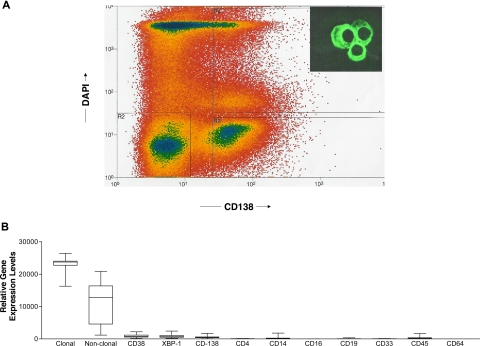
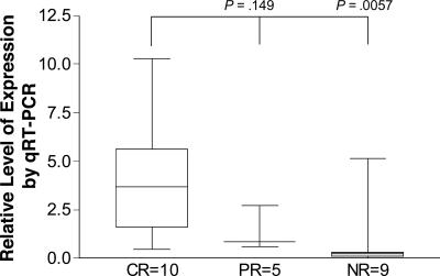
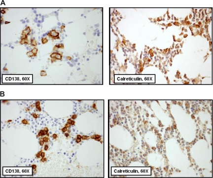
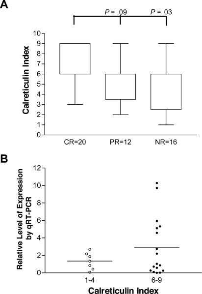
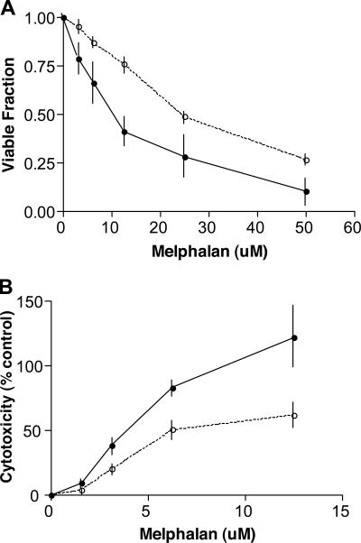
In high doses with stem-cell transplantation, melphalan is an effective but toxic therapy for patients with systemic light-chain (AL-) amyloidosis, a protein deposition and monoclonal plasma cell disease. Melphalan can eliminate the indolent clonal plasma cells that cause the disease, an achievement called a complete response. Such a response is usually associated with extended survival, while no response (a less than 50% reduction) is not. Gene-expression studies and a stringently supervised analysis identified calreticulin as having significantly higher expression in the pretreatment plasma cells of patients with systemic AL-amyloidosis who then had a complete response to high-dose melphalan. Calreticulin is a pleiotropic calcium-binding protein found in the endoplasmic reticulum and the nucleus whose overexpression is associated with increased sensitivity to apoptotic stimuli. Real-time PCR and immunohistochemical staining also showed that expression of calreticulin was higher in the plasma cells of those with a complete response. Furthermore, wild-type murine embryonic fibroblasts were significantly more sensitive to melphalan than calreticulin knock-out murine embryonic fibroblasts. These data have important implications for understanding the activity of melphalan in plasma-cell diseases and support further investigation of calreticulin and its modulation in patients with systemic AL-amyloidosis receiving high-dose melphalan.
Figures
Similar articles
-
High-dose melphalan and peripheral blood stem cell transplantation for light-chain amyloidosis with cardiac involvement.Blood. 2012 Feb 2;119(5):1117-22. doi: 10.1182/blood-2011-07-370031. Epub 2011 Dec 6. Blood. 2012. PMID: 22147893 Free PMC article.
-
High-dose melphalan and stem cell transplantation for patients with AL amyloidosis: trends in treatment-related mortality over the past 17 years at a single referral center.Blood. 2012 Nov 22;120(22):4445-6. doi: 10.1182/blood-2012-09-457341. Blood. 2012. PMID: 23175664 No abstract available.
-
High-dose melphalan versus melphalan plus dexamethasone for AL amyloidosis.N Engl J Med. 2007 Sep 13;357(11):1083-93. doi: 10.1056/NEJMoa070484. N Engl J Med. 2007. PMID: 17855669 Clinical Trial.
-
High-Dose Melphalan and Autologous Peripheral Blood Stem Cell Transplantation in AL Amyloidosis.Acta Haematol. 2020;143(4):381-387. doi: 10.1159/000506498. Epub 2020 Apr 3. Acta Haematol. 2020. PMID: 32248194 Review.
-
Recent advances in the management of AL Amyloidosis.Br J Haematol. 2016 Jan;172(2):170-86. doi: 10.1111/bjh.13805. Epub 2015 Oct 22. Br J Haematol. 2016. PMID: 26491974 Review.
Cited by
-
Heterohybridomas producing human immunoglobulin light chains using CD138-selected bone marrow cells.Biochem Biophys Rep. 2025 Apr 23;42:102017. doi: 10.1016/j.bbrep.2025.102017. eCollection 2025 Jun. Biochem Biophys Rep. 2025. PMID: 40330075 Free PMC article.
-
siRNA targeting the κ light chain constant region: preclinical testing of an approach to nonfibrillar and fibrillar light chain deposition diseases.Gene Ther. 2016 Oct;23(10):727-733. doi: 10.1038/gt.2016.50. Epub 2016 Jun 20. Gene Ther. 2016. PMID: 27383253
-
Calreticulin and cancer.Pathol Oncol Res. 2013 Apr;19(2):149-54. doi: 10.1007/s12253-012-9600-2. Epub 2013 Feb 8. Pathol Oncol Res. 2013. PMID: 23392843 Review.
-
Unfolded protein response suppresses CEBPA by induction of calreticulin in acute myeloid leukaemia.J Cell Mol Med. 2010 Jun;14(6B):1509-19. doi: 10.1111/j.1582-4934.2009.00870.x. Epub 2009 Jul 31. J Cell Mol Med. 2010. PMID: 19659458 Free PMC article.
-
Primary myeloma interaction and growth in coculture with healthy donor hematopoietic bone marrow.BMC Cancer. 2015 Nov 6;15:864. doi: 10.1186/s12885-015-1892-7. BMC Cancer. 2015. PMID: 26545722 Free PMC article.
References
-
- Pepys MB. Amyloidosis. Annu Rev Med. 2006;57:223–241. - PubMed
-
- Merlini G, Westermark P. The systemic amyloidoses: clearer understanding of the molecular mechanisms offers hope for more effective therapies. J Intern Med. 2004;255:159–178. - PubMed
-
- Merlini G, Bellotti V. Molecular mechanisms of amyloidosis. N Engl J Med. 2003;349:583–596. - PubMed
-
- Skinner M, Sanchorawala V, Seldin DC, et al. High-dose melphalan and autologous stem-cell transplantation in patients with AL amyloidosis: an 8-year study. Ann Intern Med. 2004;140:85–93. - PubMed
-
- Comenzo RL. Hematopoietic cell transplantation for primary systemic amyloidosis: what have we learned. Leuk Lymphoma. 2000;37:245–258. - PubMed
Publication types
MeSH terms
Substances
Grants and funding
LinkOut - more resources
Full Text Sources
Other Literature Sources
Medical
Research Materials
