. 2007;35(22):7665-75.
doi: 10.1093/nar/gkm933. Epub 2007 Nov 3.

Feedback-regulated poly(ADP-ribosyl)ation by PARP-1 is required for rapid response to DNA damage in living cells

Affiliations

Feedback-regulated poly(ADP-ribosyl)ation by PARP-1 is required for rapid response to DNA damage in living cells

Oliver Mortusewicz et al. Nucleic Acids Res. 2007.

Abstract

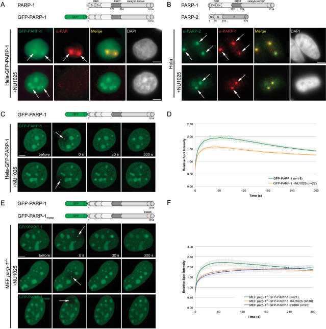
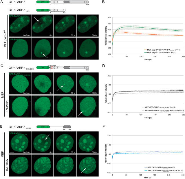
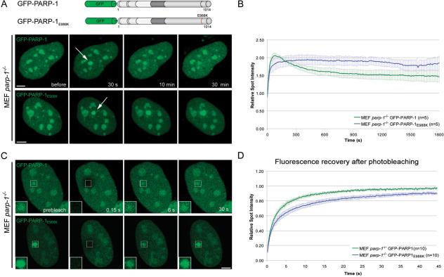
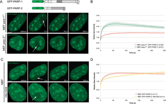
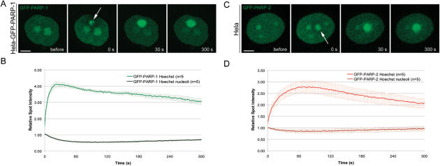
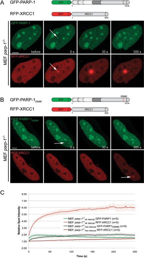
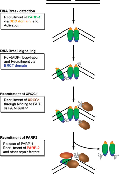
Genome integrity is constantly threatened by DNA lesions arising from numerous exogenous and endogenous sources. Survival depends on immediate recognition of these lesions and rapid recruitment of repair factors. Using laser microirradiation and live cell microscopy we found that the DNA-damage dependent poly(ADP-ribose) polymerases (PARP) PARP-1 and PARP-2 are recruited to DNA damage sites, however, with different kinetics and roles. With specific PARP inhibitors and mutations, we could show that the initial recruitment of PARP-1 is mediated by the DNA-binding domain. PARP-1 activation and localized poly(ADP-ribose) synthesis then generates binding sites for a second wave of PARP-1 recruitment and for the rapid accumulation of the loading platform XRCC1 at repair sites. Further PARP-1 poly(ADP-ribosyl)ation eventually initiates the release of PARP-1. We conclude that feedback regulated recruitment of PARP-1 and concomitant local poly(ADP-ribosyl)ation at DNA lesions amplifies a signal for rapid recruitment of repair factors enabling efficient restoration of genome integrity.


Figures

Figure 1.
Figure 1.
Recruitment of PARP-1 to DNA damage sites. (A) Immunostaining of PAR after microirradiation of Hela cells stably transfected with GFP-PARP-1. GFP-PARP-1 clearly colocalizes with PAR at microirradiated sites. Treatment of Hela GFP-PARP-1 cells with the PARP-1 inhibitor NU1025 results in loss of PAR signals at microirradiated sites, while GFP-PARP-1 accumulation is still present. (B) Immunostaining of PARP-1 and PARP-2 after microirradiation of Hela cells in the absence or presence of NU1025. (C) Live cell imaging of microirradiated Hela cells stably expressing GFP-PARP-1. Accumulation of GFP-PARP-1 can be observed immediately after microirradiation in untreated cells as well as in cells treated with the PARP inhibitor NU1025. (D) Quantitative evaluation of PARP-1 recruitment kinetics in the absence and presence of the PARP inhibitor NU1025. Inhibition of PARP activity does not prevent recruitment of PARP-1 but leads to a reduced accumulation at microirradiated sites. (E and F) Live cell imaging and quantitative evaluation of PARP-1 recruitment kinetics in the absence and presence of the PARP inhibitor NU1025 compared with the recruitment kinetics of the fluorescence tagged catalytic mutant PARP-1 after microirradiation of PARP-1 knock out cells. Error bars represent the SEM. Scale bar, 5 μm.
Figure 2.
Figure 2.
Mechanism of PARP-1 recruitment to DNA damage sites. (A) Live cell imaging of microirradiated PARP-1 knock out MEFs (MEF parp-1−/−) expressing either GFP-PARP-1 or the GFP-tagged DNA binding domain of PARP-1 (GFP-PARP-11–373). Accumulation of both, GFP-PARP-1 and GFP-PARP-11–373 can be observed immediately after microirradiation. (B) Quantitative evaluation of GFP-PARP-11–373 recruitment kinetics. For comparision, the recruitment kinetics of GFP-PARP-1 from Figure 1F are displayed. Time-matched controls are shown in Supplementary Figure 3. (C) Live cell imaging of microirradiated MEFs expressing a PARP-1 fusion protein containing two point mutations affecting the DNA binding capacities of PARP-1 (GFP-PARP-1C21G,C25G) in the absence or presence of the PARP inhibitor NU1025. (D) Quantitative evaluation of recruitment kinetics. (E) Live cell imaging of microirradiated MEFs expressing the GFP-tagged BRCT domain of PARP-1 (GFP-PARP-1384–524) in the absence or presence of the PARP inhibitor NU1025. (F) Quantitative evaluation of recruitment kinetics. Error bars represent the SEM. Scale bar, 5 μm.
Figure 3.
Figure 3.
The catalytic activity of PARP-1 is needed for dissociation from DNA damage sites. (A) Long-term observations of microirradiated PARP-1 knock out MEFs (MEF parp-1−/−) expressing either GFP-PARP-1 or a GFP-tagged catalytic mutant (GFP-PARP-1E988K). The catalytic mutant shows a prolonged association at DNA damage sites. (B) Quantitative evaluation of recruitment kinetics. (C) Mobility of GFP-PARP-1 and GFP-PARP-1E988K at DNA damage sites. The mobility of accumulated fluorescent fusion proteins was determined by bleaching the microirradiated site 5 min after microirradiation and subsequent recovery measurements. Inset shows the bleached microirradiated site. (D) FRAP data from 10 individual experiments are shown as mean curves. Error bars represent the SEM. Scale bar, 5 μm.
Figure 4.
Figure 4.
Recruitment of PARP-2 to DNA damage sites in living cells. (A) Live cell imaging of microirradiated MEFs either expressing GFP-PARP-1 or GFP-PARP-2. Accumulation of GFP-PARP-1 and GFP-PARP-2 can be observed immediately after microirradiation. (B) Quantitative evaluation of GFP-PARP-2 recruitment kinetics. For comparision, the recruitment kinetics of GFP-PARP-1 from Figure 1F are displayed. Time-matched controls are shown in Supplementary Figure 3. (C and D) Live cell imaging of microirradiated MEFs reveals a slower accumulation of GFP-PARP-2 in the presence of NU1025. Error bars represent the SEM. Scale bar, 5 μm.
Figure 5.
Figure 5.
The Nucleolus serves as a storage of PARP-1 and PARP-2 to cope with heavy DNA damage. (A and C) Live cell imaging of microirradiated Hela cells sensitized with Hoechst 33285. Microirradiation of Hoechst sensitized cells leads to massive recruitment and temporary depletion of PARP-1 and PARP-2 from the nucleolus. (B and D) Quantitative evaluation of recruitment and nucleolar depletion kinetics. Error bars represent the SEM. Scale bar, 5 μm.
Figure 6.
Figure 6.
Efficient recruitment of XRCC1 to DNA repair sites depends on the presence of PARP-1. (A) Live cell imaging of microirradiated wild-type, PARP-1 and PARP-2 knock out MEFs (MEF parp-1−/−, MEF parp-2−/−) expressing GFP-XRCC1. Accumulation of GFP-XRCC1 at DNA damage sites is dramatically reduced in the absence of PARP-1. (B) Quantitative evaluation of recruitment kinetics. (C and D) Mobility of GFP-XRCC1 at DNA damage sites. The mobility of accumulated fluorescent fusion proteins was determined by bleaching the microirradiated site 5 min after microirradiation and subsequent recovery measurements. Inset shows the bleached microirradiated site. FRAP data from 10 individual experiments are shown as mean curves. Error bars represent the SEM. Scale bar, 5 μm.
Figure 7.
Figure 7.
The catalytic activity of PARP-1 is needed for efficient recruitment of XRCC1 to laser-induced DNA damage sites. (A) Live cell imaging of microirradiated PARP-1 knock out MEFs (MEF parp-1−/−) coexpressing GFP-PARP-1 and RFP-XRCC1. Expression of GFP-tagged wild-type PARP-1 results in efficient recruitment of RFP-XRCC1. (B) Live cell imaging of microirradiated PARP-1 knock out MEFs (MEF parp-1−/−) coexpressing GFP-PARP-1E988K and RFP-XRCC1. Accumulation of RFP-XRCC1 at DNA damage sites is dramatically reduced in PARP-1 knock out MEFs expressing catalytically inactive GFP-PARP-1E988K. (C) Quantitative evaluation of recruitment kinetics. Error bars represent the SEM. Scale bar, 5 μm.
Figure 8.
Figure 8.
Simplified model for the recruitment of repair factors to SSB. See text for a detailed discussion of the role and regulation of PARPs.

References

    1. Hoeijmakers JH. Genome maintenance mechanisms for preventing cancer. Nature. 2001;411:366–374. - PubMed
    1. Schreiber V, Dantzer F, Ame JC, de Murcia G. Poly(ADP-ribose): novel functions for an old molecule. Nat. Rev. Mol. Cell Biol. 2006;7:517–528. - PubMed
    1. Mackey ZB, Niedergang C, Murcia JM, Leppard J, Au K, Chen J, de Murcia G, Tomkinson AE. DNA ligase III is recruited to DNA strand breaks by a zinc finger motif homologous to that of poly(ADP-ribose) polymerase. Identification of two functionally distinct DNA binding regions within DNA ligase III. J. Biol. Chem. 1999;274:21679–21687. - PubMed
    1. Gradwohl G, Menissier de Murcia JM, Molinete M, Simonin F, Koken M, Hoeijmakers JH, de Murcia G. The second zinc-finger domain of poly(ADP-ribose) polymerase determines specificity for single-stranded breaks in DNA. Proc. Natl Acad. Sci. USA. 1990;87:2990–2994. - PMC - PubMed
    1. Pleschke JM, Kleczkowska HE, Strohm M, Althaus FR. Poly(ADP-ribose) binds to specific domains in DNA damage checkpoint proteins. J. Biol. Chem. 2000;275:40974–40980. - PubMed

Publication types