

Fatal pancreatic panniculitis associated with acute pancreatitis: a case report
- PMID: 17982246
- PMCID: PMC2693864
- DOI: 10.3346/jkms.2007.22.5.914
Fatal pancreatic panniculitis associated with acute pancreatitis: a case report
Abstract
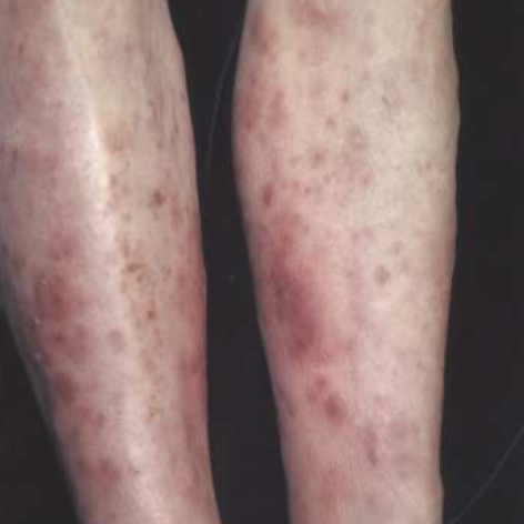
Pancreatic panniculitis is a rare disease in which necrosis of fat in the panniculus and other distant foci occurs in the setting of pancreatic diseases; these diseases include acute and chronic pancreatitis, pancreatic carcinoma, pseudocyst, and other pancreatic diseases. This malady is manifested as tender erythematous nodules on the legs, buttock, or trunk. Histopathologically, it shows the pathognomonic findings of focal subcutaneous fat necrosis and ghost-like anucleated cells with a thick shadowy wall. We herein report a case of fatal pancreatic panniculitis that was associated with acute pancreatitis in a 50-yr-old man. He presented with a 3-week history of multiple tender skin nodules, abdominal pain and distension. Laboratory and radiologic findings revealed acute pancreatitis, and skin biopsy showed pancreatic panniculitis. Despite intensive medical care, he died of multi-organ failure 3 weeks after presentation.
Figures


Similar articles
-
Acute septal panniculitis. A cutaneous marker of a very early stage of pancreatic panniculitis indicating acute pancreatitis.JOP. 2005 Jul 8;6(4):334-8. JOP. 2005. PMID: 16006683
-
[Pancreatic Panniculitis in Patients with Chronic Pancreatitis: Case Report and Review of Literature].Korean J Gastroenterol. 2017 Jan 25;69(1):83-86. doi: 10.4166/kjg.2017.69.1.83. Korean J Gastroenterol. 2017. PMID: 28135797 Review. Korean.
-
Subcutaneous fat necrosis/panniculitis and polyarthritis associated with acinar cell carcinoma of the pancreas: a rare presentation of pancreatitis, panniculitis and polyarthritis syndrome.J Drugs Dermatol. 2010 Sep;9(9):1145-50. J Drugs Dermatol. 2010. PMID: 20865849
-
A case report of pancreatic panniculitis due to acute pancreatitis with intraductal papillary mucinous neoplasm.BMC Gastroenterol. 2020 Aug 24;20(1):286. doi: 10.1186/s12876-020-01430-9. BMC Gastroenterol. 2020. PMID: 32831035 Free PMC article.
-
Pancreatic panniculitis.G Ital Dermatol Venereol. 2013 Aug;148(4):419-25. G Ital Dermatol Venereol. 2013. PMID: 23900163 Review.
Cited by
-
[Pancreatic panniculitis with multiple osteolytic lesions].Hautarzt. 2015 Feb;66(2):114-6. doi: 10.1007/s00105-014-3514-8. Hautarzt. 2015. PMID: 25325989 German.
-
Risk of Pancreatic Cancer After Acute Pancreatitis: A Retrospective Analysis of the Korean National Sample Cohort.J Korean Med Sci. 2024 Jan 29;39(4):e21. doi: 10.3346/jkms.2024.39.e21. J Korean Med Sci. 2024. PMID: 38288535 Free PMC article.
-
Panniculitis - A Rare Manifestation of Acute Pancreatitis.GE Port J Gastroenterol. 2015 Mar 29;22(3):117-120. doi: 10.1016/j.jpge.2015.01.007. eCollection 2015 May-Jun. GE Port J Gastroenterol. 2015. PMID: 28868388 Free PMC article.
-
Skin manifestations of pancreatic diseases.Biomed Pap Med Fac Univ Palacky Olomouc Czech Repub. 2022 Dec;166(4):353-358. doi: 10.5507/bp.2022.035. Epub 2022 Jul 22. Biomed Pap Med Fac Univ Palacky Olomouc Czech Repub. 2022. PMID: 35938387 Review.
-
[Multiple pressure-sensitive, ulcerating nodes on both lower legs of a 84-year-old woman].Hautarzt. 2015 Jan;66(1):74-6. doi: 10.1007/s00105-014-3535-3. Hautarzt. 2015. PMID: 25392127 German. No abstract available.
References
-
- Oh YS, Kang BD, Kim IH, Kye YC, Kim SN. Case reports: a case of subcutaneous fat necrosis associated with pancreatitis. Ann Dermatol. 1996;8:38–42.
-
- Johnson MA, Kannan DG, Balachandar TG, Jeswanth S, Rajendran S, Surendran R. Acute septal panniculitis. A cutaneous marker of a very early stage of pancreatic panniculitis indicating acute pancreatitis. JOP. 2005;6:334–338. - PubMed
-
- Kim HJ, Lee KG. Subcutaneous fat necrosis associated with pancreatic adenocarcinoma: a case report. Korean J Pathol. 1996;30:155–160.
-
- Park SW, Wang HY, Lee HJ, Wang JK. A case of nodular fat necrosis associated with pancreatitis. a case report. Korean J Dermatol. 1998;36:346–349.
-
- Mullin GT, Caperton EM, Jr, Crespin SR, Williams RC., Jr Arthritis and skin lesions resembling erythema nodosum in pancreatic disease. Ann Intern Med. 1968;68:75–87. - PubMed
Publication types
MeSH terms
LinkOut - more resources
Full Text Sources
Medical
