. 2007 Nov 7;2(11):e1155.
doi: 10.1371/journal.pone.0001155.

Common activation of canonical Wnt signaling in pancreatic adenocarcinoma

Affiliations

Common activation of canonical Wnt signaling in pancreatic adenocarcinoma

Marina Pasca di Magliano et al. PLoS One. .

Abstract

Pancreatic ductal adenocarcinoma (PDA) is an extremely aggressive malignancy, which carries a dismal prognosis. Activating mutations of the Kras gene are common to the vast majority of human PDA. In addition, recent studies have demonstrated that embryonic signaling pathway such as Hedgehog and Notch are inappropriately upregulated in this disease. The role of another embryonic signaling pathway, namely the canonical Wnt cascade, is still controversial. Here, we use gene array analysis as a platform to demonstrate general activation of the canonical arm of the Wnt pathway in human PDA. Furthermore, we provide evidence for Wnt activation in mouse models of pancreatic cancer. Our results also indicate that Wnt signaling might be activated downstream of Hedgehog signaling, which is an early event in PDA evolution. Wnt inhibition blocked proliferation and induced apoptosis of cultured adenocarcinoma cells, thereby providing evidence to support the development of novel therapeutical strategies for Wnt inhibition in pancreatic adenocarcinoma.


Conflict of interest statement

Competing Interests: The authors have declared that no competing interests exist.

Figures

Figure 1
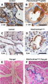
Figure 1. Active Wnt signaling in a mouse model of PDA.
A. Immunostaning for β-Catenin in a control pancreas is confined to the plasma membrane. B. In contrast, cytoplasmic β-catenin expression is observed in PanIN lesions and tumor of a 8-week old Pdx-Cre;KrasG12D;p53f/+ pancreas. The tumor is outlined in red. Note the accumulation of β-catenin in the epithelial cells (brown staining). Two PanIN lesions are outlined in light blue. The black arrow points at cells that have retained membranous β-catenin, while the red arrows point at areas of accumulation of β-catenin in the cytoplasm. C. Tcf4 is expressed at a low level in ductal cells in a wild-type mouse pancreas. D. Elevated expression of Tcf4 in PanIN lesions of a 12-week old Pdx-Cre;KrasG12D pancreas. E, F. LacZ staining of pancreatic tissue of adult (6 months old) Top-gal (E) and Pdx-Cre;KrasG12D;Top-gal (F) mice. No staining is present in the Top-gal pancreas. ß-galactosidase activity is detected in the PanIN lesions of the Pdx-Cre;KrasG12D;Top-gal pancreas, indicating activation of the canonical Wnt pathway. The black bar represents 35 µm in A-D, 67.5 µm in E, F.
Figure 2
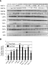
Figure 2. Wnt signaling is active in human pancreatic adenocarcinoma cell lines.
A. RT-PCR analysis for components of the Wnt signaling pathway in 26 human pancreatic adenocarcinoma cell lines: 1-MiaPaca2; 2-Panc1; 3-CFPAC1; 4-HPAFII; 5-Capan-2; 6-AsPC1; 7-Hs766T; 8-BxPC3; 9-COLO357; 10-L3.3; 11-L3.6sl; 12-L3.6pl; 13-SW1990; 14-SU86.86; 15-PL45; 16-HPAC; 17-MPanc96; 18-Panc1.28; 19-Panc2.03; 20-Panc2.13; 21-Panc3.27; 22-Panc4.21; 23-Panc5.04; 24-Panc6.03; 25-Panc8.13; 26-Panc10.05. B. Wnt signaling is active in all nine pancreatic adenocarcinoma cell lines tested, as indicated by the activation of the Top-Flash reporter (black) compared to the basal activity of the Fop-Flash reporter (white). Renilla luciferase was used to normalize for transfection efficiency. Error bars are shown as St. Dev. P-values were calculated in comparison to control Fop-Flash activity (white bars). *, p<0.05; **, p<0.01.
Figure 3
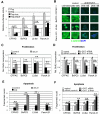
Figure 3. Inhibition of Wnt signaling blocks adenocarcinoma cell proliferation.
A. Inhibition of Wnt signaling in four pancreatic adenocarcinoma cell lines. Cells transfected with Fop-Flash (white) or Top-Flash (light gray) reporter constructs were co-transfected with an empty expression vector or an expression vector containing the Wnt-inhibitors Icat (dark gray) or dominant-negative Lef1 (dnLef; black). Fop-Flash activity was not affected by either Icat or dn-Lef co-transfection, thus only one Fop-Flash data point is shown. The significant reduction of Top-Flash activity in cells expressing the inhibitors indicates inhibition of Wnt signaling in pancreatic cancer cells. P-values were calculated in comparison to Top-Flash activity (light gray bar). B. Immunofluorescent staining against β-CATENIN in four pancreatic adenocarcinoma cell lines after transfection with a siRNA directed against β-CATENIN. The β-CATENIN protein levels are dramatically decreased in a dose-dependent manner following β-CATENIN siRNA transfection. Control siRNA transfected cells have β-CATENIN levels indistinguishable from those found in untransfected cells (untreated). C. Transfection with an Icat–IRES-eGFP expression vector (Icat, black) or dominant negative Lef – IRES-eGFP (dnLef, gray) strongly inhibits growth of four pancreatic cancer cells lines, measured as the ability to incorporate BrdU. Control cells (white) were transfected with the IRES-eGFP expression vector; all cells were harvested 48 hrs after transfection. P-values are shown in comparison to control transfected cells. D. Proliferation is reduced in a dose-dependent manner in cells treated for 48hrs with anti-ß-CATENIN siRNA (dark grey, black,) compared to control cells (white). Untreated (no siRNA), white columns; control siRNA (300 nM), light grey columns; anti-ß-CATENIN siRNA (100 nM), dark grey columns; anti-ß-CATENIN siRNA (300 nM), black columns. P-values are shown in comparison to untreated controls (white columns). E. Level of apoptosis, measured as cells with DNA content lower than the diploid amount, in control transfected cells (white) or cells transfected with Icat (black) or dominant negative Lef (grey). F. Treatment with anti-ß-CATENIN siRNA increases levels of apoptosis in a concentration-dependent manner. Error bars are shown as St. Dev.; P-values, #, not significant; *, p<0.05; **, p<0.01.
Figure 4
Figure 4. Hedgehog signaling regulates Wnt activity in untransformed duct cells.
A. Activity of the Wnt and Hedgehog pathways in control PDCs and stable clones transfected with dominant-active forms of GLI2 (PDC-GLI2). Activation of the Hedgehog pathway in two PDC-GLI2 clones (#1, #2) results in significant increase in Wnt signaling activity. B. Activity of the Wnt and Hedgehog pathways in control PDCs and PDCs stably transfected with a dominant active form of Lef1 (PDC-Lef–da). Activation of the Wnt pathway in the PDC-Lef–da cells does not affect the level of Hedgehog signaling. Error bars are shown as St. Dev. P-values, #, not significant; **p<0.01. C. PDC cells were stably transfected with a myc-tagged version of the constitutive active form of GLI2 (GLI2-myc fusion protein). Immunostaining using an anti-myc antibody confirms GLI2 expression in transfected cells. Untransfected PDC cells are shown as negative control. Immunostaining for Tcf4 is not detectable in wt PDC cells, but it is strongly upregulated in PDC-GLI2 cells. D. Immunohistochemistry analysis of Panc4.21 and L3.6sl pancreatic cancer cell lines shows that inhibition of the Wnt signaling pathway using anti-β-CATENIN siRNA (100nM) results in a strong downregulation of TCF4 expression compared to control cells (transfected with an unrelated siRNA). Similarly, inhibition of the Hedgehog pathway using an anti-GLI1/2 siRNA (100 nM) results in TCF4 downregulation; the same effect is observed in cells treated with a combination of anti-β-CATENIN and anti-GLI1/2 siRNA (each 100nM). E. Quantification of Tcf4 expression in PDC (white) and PDC-GLI2 (black) cells. At least 100 cell nuclei were scored for Tcf4 expression and the percentage of positive nuclei is shown in the histogram. F. Quantification of TCF4 positive nuclei following siRNA treatment in human PDA cell lines. At least 100 nuclei were scored for each category: control siRNA (white), anti-β-CATENIN siRNA (light gray), anti-GLI siRNA (dark gray) and a combination of both siRNAs (black). Error bars are shown as St. Dev.; p-values #, not significant; *p<0.05; **, p<0.01. G. In wild-type mouse pancreas tissue, β-catenin is only localized at the cell membrane. H. Presence of nuclear β-catenin in a fraction of the tumors cells in an undifferentiated pancreatic tumor of a Pdx-Cre;CLEG2 mouse. I. Cytoplasmic and nuclear β-catenin staining in the PanIN lesions of a Pdx-Cre;CLEG2;KrasG12D pancreas. The black bar represents 35 µm in G-I.

References

    1. Hezel AF, Kimmelman AC, Stanger BZ, Bardeesy N, Depinho RA. Genetics and biology of pancreatic ductal adenocarcinoma. Genes Dev. 2006;20:1218–1249. - PubMed
    1. Hruban RH, Takaori K, Klimstra DS, Adsay NV, Albores-Saavedra J, et al. An illustrated consensus on the classification of pancreatic intraepithelial neoplasia and intraductal papillary mucinous neoplasms. Am J Surg Pathol. 2004;28:977–987. - PubMed
    1. Biankin AV, Kench JG, Dijkman FP, Biankin SA, Henshall SM. Molecular pathogenesis of precursor lesions of pancreatic ductal adenocarcinoma. Pathology. 2003;35:14–24. - PubMed
    1. Biankin AV, Kench JG, Morey AL, Lee CS, Biankin SA, et al. Overexpression of p21(WAF1/CIP1) is an early event in the development of pancreatic intraepithelial neoplasia. Cancer Res. 2001;61:8830–8837. - PubMed
    1. Miyamoto Y, Maitra A, Ghosh B, Zechner U, Argani P, et al. Notch mediates TGF alpha-induced changes in epithelial differentiation during pancreatic tumorigenesis. Cancer Cell. 2003;3:565–576. - PubMed

Publication types