. 2007 Nov;3(11):e161.
doi: 10.1371/journal.ppat.0030161.

Macrophage activation redirects yersinia-infected host cell death from apoptosis to caspase-1-dependent pyroptosis

Affiliations

Macrophage activation redirects yersinia-infected host cell death from apoptosis to caspase-1-dependent pyroptosis

Tessa Bergsbaken et al. PLoS Pathog. 2007 Nov.

Abstract

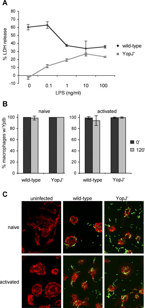
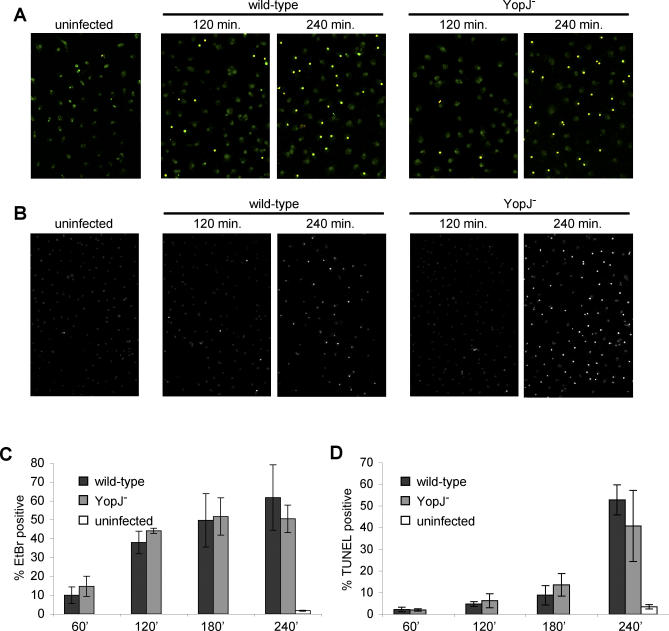
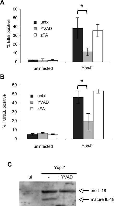
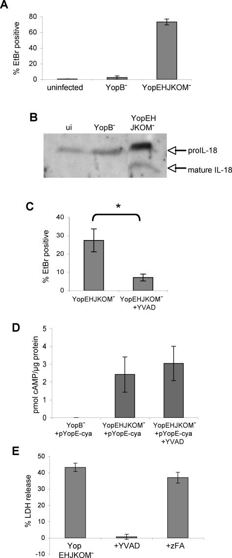
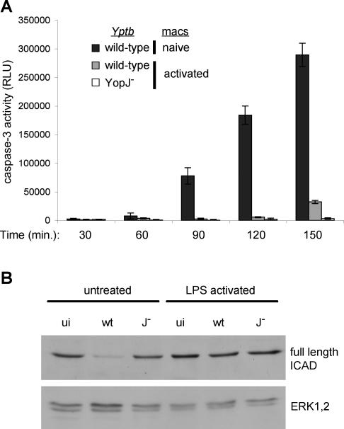
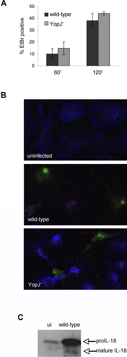
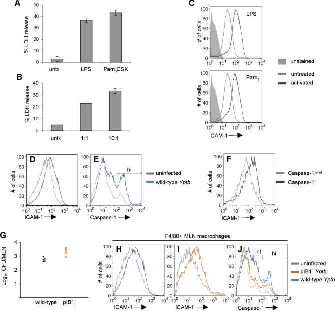
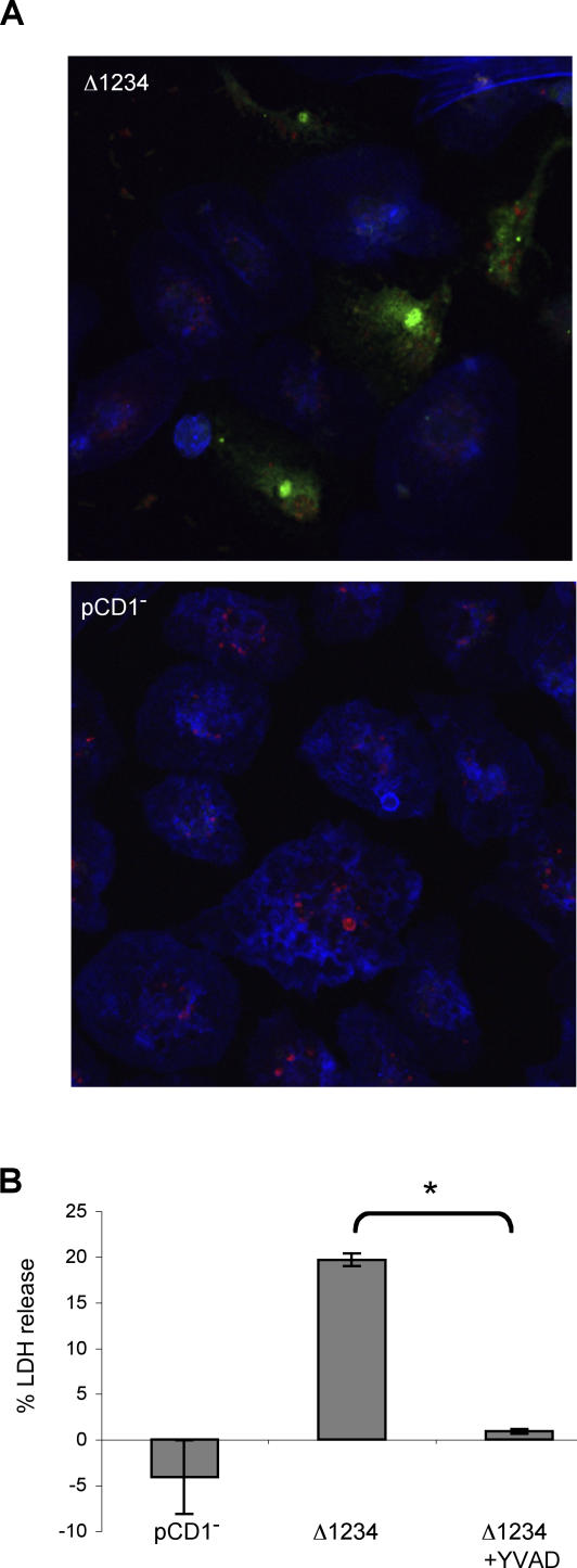
Infection of macrophages by Yersinia species results in YopJ-dependent apoptosis, and naïve macrophages are highly susceptible to this form of cell death. Previous studies have demonstrated that macrophages activated with lipopolysaccharide (LPS) prior to infection are resistant to YopJ-dependent cell death; we found this simultaneously renders macrophages susceptible to killing by YopJ(-) Yersinia pseudotuberculosis (Yptb). YopJ(-) Yptb-induced macrophage death was dependent on caspase-1 activation, resulting in rapid permeability to small molecules, followed by membrane breakdown and DNA damage, and accompanied by cleavage and release of proinflammatory interleukin-18. Induction of caspase-1-dependent death, or pyroptosis, required the bacterial type III translocon but none of its known translocated proteins. Wild-type Yptb infection also triggered pyroptosis: YopJ-dependent activation of proapoptotic caspase-3 was significantly delayed in activated macrophages and resulted in caspase-1-dependent pyroptosis. The transition to susceptibility was not limited to LPS activation; it was also seen in macrophages activated with other Toll-like receptor (TLR) ligands and intact nonviable bacteria. Yptb infection triggered macrophage activation and activation of caspase-1 in vivo. Y. pestis infection of activated macrophages also stimulated caspase-1 activation. These results indicate that host signaling triggered by TLR and other activating ligands during the course of Yersinia infection redirects both the mechanism of host cell death and the downstream consequences of death by shifting from noninflammatory apoptosis to inflammatory pyroptosis.


Conflict of interest statement

Competing interests. The authors have declared that no competing interests exist.

Figures

Figure 1
Figure 1. Activation of Macrophages Increases Susceptibility to Cell Death Induced by Infection with YopJ Yptb
BMDMs were treated with varying concentrations of LPS for 18 h prior to infection with wild-type or YopJ Yptb. (A) Host cell lysis was assessed by measuring release of cytosolic LDH into the supernatant at 3.5 h postinfection. Data shown are means and SDs calculated from three replicates and are representative of three experiments. (B and C) Naïve macrophages and macrophages activated with 100 ng/ml LPS for 18 h were infected with GFP-expressing wild-type or YopJ Yptb. The uniformity of host cell infection was assessed by confocal microscopy. The percentage of macrophages with associated Yptb immediately after (black bars) and 120 min (gray bars) postinfection was determined (B). Means and SDs were calculated from more than five fields with a minimum of 150 cells for each condition. GFP-expressing Yptb are shown interacting with host cells at 120 min postinfection (C). Host cells were visualized by staining actin with Texas Red-phalloidin; representative images are shown.
Figure 2
Figure 2. Wild-Type and YopJ Yptb Infection of Activated Macrophages Stimulates Membrane Permeabilization Followed by DNA Damage
The kinetics of cell death were examined in LPS-activated BMDMs infected with wild-type and YopJ Yptb. (A) Macrophages labeled with SYTO10 (green) were stained with membrane-impermeant EtBr (MW = 394 Da, red) and examined by confocal microscopy to assess increases in membrane permeability (EtBr-positive/SYTO10-labeled, yellow). Representative images are shown. (B) DNA damage was also assessed by TUNEL and confocal microscopy in the same experiment. Representative images are shown. (C) The percentage of EtBr-positive/SYTO10-labeled cells was determined; data shown are means and SDs from four or more fields with a minimum of 350 cells total for each time point. Results shown are representative of two experiments. (D) The percentage of TUNEL-positive cells was determined; data shown are means and SDs from four or more fields with a minimum of 450 cells total for each time point. Results shown are representative of two experiments.
Figure 3
Figure 3. YopJ Yptb-Induced Membrane Permeability and DNA Damage Are Caspase-1-Dependent and Accompanied by Inflammatory Cytokine Processing
LPS-activated BMDMs were treated with caspase-1 inhibitor (YVAD) or negative control (zFA) peptide (200 μM) during infection with YopJ Yptb. Membrane permeability was examined by SYTO10/EtBr staining (see Figure 2A legend) and confocal microscopy at 120 min postinfection (A). DNA damage was assessed using TUNEL and confocal microscopy at 240 min postinfection (B). The percentage of EtBr/TUNEL-positive cells was determined from four or more fields with a minimum of 1,000 cells total for each condition. Representative of two experiments. * p < 0.0005. (C) Western blot analysis of mature IL-18 released into the supernatant by activated macrophages at 90 min postinfection with YopJ Yptb confirms caspase-1 activation and cytokine processing. Representative of two experiments. ui, uninfected.
Figure 4
Figure 4. Yptb-Induced Pyroptosis Is T3SS-Dependent
LPS-activated BMDMs were infected with YopEHJKOM (T3SS+, type III effector) or YopB (T3SS) Yptb. (A) Membrane permeability was examined at 90 min postinfection by EtBr/SYTO10 staining (See Figure 2A legend) and confocal microscopy. Data shown are from four or more fields with a minimum of 1,000 cells total for each condition. Representative of two experiments. (B) Western blot analysis of mature IL-18 released into the supernatant by infected macrophages at 90 min postinfection confirms caspase-1 activation. Representative of two experiments. ui, uninfected. (C) Membrane permeability was examined in infected macrophages treated with YVAD by SYTO10/EtBr staining and confocal microscopy at 45 min postinfection. Data shown are from four or more fields with a minimum of 1,000 cells for each condition. Representative of three experiments. * p < 0.0001. (D) Translocation of a YopE-adenylate cyclase fusion protein into the cytoplasm of activated macrophages was assessed by quantifying cAMP levels at 60 min postinfection, and demonstrated equivalent levels of effector translocation by YopEHJKOM Yptb in the presence of YVAD. Data shown are means and SDs calculated from three replicates. (E) Membrane breakdown and LDH release from macrophages treated with YVAD or negative control peptide zFA 120 min postinfection with YopEHJKOM Yptb. Data shown are means and SDs calculated from three replicates and are representative of three experiments.
Figure 5
Figure 5. Macrophage Activation Antagonizes Caspase-3 Activity and Apoptosis during Infection with Wild-Type Yptb
Untreated and LPS-activated BMDMs were infected with wild-type or YopJ Yptb. (A) Kinetics of caspase-3 activation in infected macrophages. Data shown are means and SDs calculated from three replicates and presented as relative light units (RLU) in cell lysates from infected samples minus uninfected controls. Representative of three experiments. At 150 min postinfection, caspase-3 activity in naïve macrophages undergoing YopJ-dependent apoptosis during infection with wild-type Yptb (289,340 ± 20,466 RLU) is greatly reduced in activated macrophages infected with wild-type Yptb (32,315 ± 2,881 RLU). Activated macrophages infected with YopJ− Yptb fail to activate caspase-3 (3,231 ± 1,531 RLU). (B) Cleavage of the caspase-3 substrate ICAD was examined in uninfected macrophages (ui) and at 120 min postinfection with wild-type (wt) or YopJ (J) Yptb by Western blot and confirmed the absence of caspase-3 activity in activated macrophages. ERK1/2 was used as a loading control.
Figure 6
Figure 6. Wild-Type Yptb Infection Induces Pyroptosis of Activated Macrophages
LPS-activated BMDMs were infected with wild-type or YopJ Yptb. (A) The kinetics of membrane permeability were examined by EtBr/SYTO10 staining (see Figure 2B legend) and confocal microscopy. Data shown are from four or more fields with a minimum of 350 cells for each time point. Representative of three experiments. (B) Macrophages were stained with FAM-YVAD (green) to identify cells with active caspase-1, Alexa633-phalloidin to visualize actin (blue), and anti-Yersinia antibodies (red) 90 min postinfection and examined by confocal microscopy. Representative images are shown. (C) Western blot analysis of mature IL-18 released into the supernatant at 90 min postinfection confirmed caspase-1 activation by wild-type Yptb infection. Representative of two experiments. ui, uninfected.
Figure 7
Figure 7. Macrophages Become Activated and Susceptible to Pyroptosis In Vivo during Murine Infection with Yptb
(A and B) BMDMs were treated with 100 ng/ml LPS, 100 ng/ml Pam3CSK4 (A), or heat-killed Yptb at ratios of 10:1 or 1:1 Yptb:macrophage (B) for 18 h and infected with YopJ Yptb. LDH release was measured after 3.5 h. Data shown are from three replicates and representative of three experiments. (C) Macrophages were activated as in (A) and surface ICAM-1 expression was measured by flow cytometry. (D–F) At 4–6 d postinfection, activated splenic macrophages from wild-type Yptb-infected mice were identified with anti-F4/80, and ICAM-1 expression and caspase-1 activity (FAM-YVAD) were measured by flow cytometry. Representative histograms of macrophage ICAM-1 expression (D), caspase-1 activity (E), and ICAM-1 expression of the caspase-1hi and caspase-1lo-int (F) macrophage populations from (E). (G–J) Mice were infected with wild-type or pIB1 Yptb and tissues were examined 5 d post-infection. CFU in the MLNs were quantified (G). Macrophages from the MLNs were identified with anti-F4/80, and ICAM-1 expression and caspase-1 activity were examined by flow cytometry. Representative histograms of MLN macrophage ICAM-1 expression from wild-type Yptb-infected (H) and pIB1 Yptb-infected (I) mice; caspase-1 activity (J).
Figure 8
Figure 8. Y. pestis Infection Induces Pyroptosis in Activated Macrophages
LPS-activated BMDMs were infected with Y. pestis Δ1234 (T3SS+, type III effector) or pCD1 (T3SS). (A) Macrophages were stained with FAM-YVAD (green) to identify cells with active caspase-1, Alexa633-phalloidin to visualize actin (blue), and anti-Yersinia antibodies (red) 90 min postinfection and examined by confocal microscopy. Representative of two experiments. (B) Membrane breakdown and LDH release from infected macrophages was inhibited by YVAD. Data shown are from three replicates and representative of two experiments. * p < 0.0001.

References

    1. Cornelis GR, Boland A, Boyd AP, Geuijen C, Iriarte M, et al. The virulence plasmid of Yersinia, an antihost genome. Microbiol Mol Biol Rev. 1998;62:1315–1352. - PMC - PubMed
    1. Cornelis GR. Yersinia type III secretion: send in the effectors. J Cell Biol. 2002;158:401–408. - PMC - PubMed
    1. Ruckdeschel K, Roggenkamp A, Lafont V, Mangeat P, Heesemann J, et al. Interaction of Yersinia enterocolitica with macrophages leads to macrophage cell death through apoptosis. Infect Immun. 1997;65:4813–4821. - PMC - PubMed
    1. Monack DM, Mecsas J, Ghori N, Falkow S. Yersinia signals macrophages to undergo apoptosis and YopJ is necessary for this cell death. Proc Natl Acad Sci U S A. 1997;94:10385–10390. - PMC - PubMed
    1. Zhang Y, Bliska JB. Immunol Role of macrophage apoptosis in the pathogenesis of Yersinia. Curr Top Microbiol. 2005;289:151–173. - PubMed

Publication types