. 2008 Apr;123(4):528-37.
doi: 10.1111/j.1365-2567.2007.02722.x. Epub 2007 Nov 5.

Innate immunity in insects: surface-associated dopa decarboxylase-dependent pathways regulate phagocytosis, nodulation and melanization in medfly haemocytes

Affiliations

Innate immunity in insects: surface-associated dopa decarboxylase-dependent pathways regulate phagocytosis, nodulation and melanization in medfly haemocytes

Maria Sideri et al. Immunology. 2008 Apr.

Abstract

Phagocytosis, melanization and nodulation in insects depend on phenoloxidase (PO) activity. In this report, we demonstrated that these three processes appear to be also dependent on dopa decarboxylase (Ddc) activity. Using flow cytometry, RNA interference, immunoprecipitation and immunofluorescence, we demonstrated the constitutive expression of Ddc and its strong association with the haemocyte surface, in the medfly Ceratitis capitata. In addition, we showed that Escherichia coli phagocytosis is markedly blocked by small interfering RNA (siRNA) for Ddc, antibodies against Ddc, as well as by inhibitors of Ddc activity, namely carbidopa and benzerazide, convincingly revealing the involvement of Ddc activity in phagocytosis. By contrast, latex beads and lipopolysaccharide (LPS) did not require Ddc activity for their uptake. It was also shown that nodulation and melanization processes depend on Ddc activation, because antibodies against Ddc and inhibitors of Ddc activity prevent haemocyte aggregation and melanization in the presence of excess E. coli. Therefore, phagocytosis, melanization and nodulation depend on haemocyte-surface-associated PO and Ddc. These three unrelated mechanisms are based on tyrosine metabolism and share a number of substrates and enzymes; however, they appear to be distinct. Phagocytosis and nodulation depend on dopamine-derived metabolite(s), not including the eumelanin pathway, whereas melanization depends exclusively on the eumelanin pathway. It must also be underlined that melanization is not a prerequisite for phagocytosis or nodulation. To our knowledge, the involvement of Ddc, as well as dopa and its metabolites, are novel aspects in the phagocytosis of medfly haemocytes.


Figures
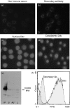
Figure 1
Figure 1
Demonstration of surface dopa decarboxylase (Ddc) on medfly haemocytes. Ddc was identified on the haemocyte surface (c) and inside haemocytes (d) with indirect immunocytofluorescent staining. Control staining was performed with either non-immune serum (a) or secondary antibody (b) to detect a non-specific signal. Ddc was also identified in haemocyte protein crude extract by immunoprecipitation and immunoblot analysis (e) and on the surface of haemocytes by flow cytometry (f), using specific antibodies. Ab”, non-specific immunoprecipitation caused by the secondary antibody; E, endogenous fluorescence; IP, immunoprecipitation; FITC, fluorescein isothiocyanate; L, haemocyte lysate; MW, molecular weight; S, supernatant after precipitation.
Figure 2
Figure 2
Developmental profile of dopa decarboxylase (Ddc) at the third instar larval stage and its stable association with the cell membrane. Flow cytometry analysis revealed the presence of Ddc on the haemocyte surface at the wandering stage, followed by a peak at the white pupa and a decrease 2 hr later (a) Indirectly fluorescein isothiocyanate (FITC)-labelled Ddc remains the same on the haemocyte surface after incubation with 0·6 m NaCl (b) AFU, arbitrary fluorescent units = (median sample fluorescence value) − (median endogenous fluorescence value).
Figure 3
Figure 3
Silencing of dopa decarboxylase (Ddc) with small interfering RNA (siRNA). Haemocytes were incubated for 7 hr with or without siRNA for Ddc. Flow cytometry showed a 90 ± 4% reduction of surface Ddc using specific antibodies (a) A control experiment was performed with an irrelevant siRNA and showed no reduction of surface Ddc (b) Non-specific binding of the secondary antibody appeared to be not significant. FITC, fluorescein isothiocyanate.
Figure 4
Figure 4
Dopa decarboxylase (Ddc) is necessary for the phagocytosis of Escherichia coli. Haemocytes were incubated with fluorescein isothiocyanate (FITC) conjugated to E. coli, lipopolysaccharide (LPS) or latex beads, with or without pre-incubation with Ddc small interfering RNA (siRNA) or anti-Ddc. Ddc siRNA and anti-Ddc reduced the phagocytosis of E. coli to about 68% and 40%, respectively, but they did not influence the uptake of LPS or latex beads. A control experiment with an irrelevant siRNA showed no influence on haemocyte phagocytosis potential. The percentages represent the mean value of three experiments.
Figure 5
Figure 5
Dopa decarboxylase (Ddc) activity is necessary for the phagocytosis of Escherichia coli. Haemocytes were incubated with fluorescein isothiocyanate (FITC) conjugated to E. coli, lipopolysaccharide (LPS) or latex beads, with or without pre-incubation with the specific Ddc inhibitors, carbidopa or benserazide. Both inhibitors reduced the phagocytosis of E. coli to ≈ 61–65% but they did not influence the uptake of LPS or latex beads. The percentages represent the mean value of three experiments.
Figure 6
Figure 6
There is no functional relationship between focal adhesion kinase (FAK) and dopa decarboxylase (Ddc). Haemocytes were incubated in Grace's medium with Escherichia coli. Haemocytes were pretreated with either Ddc small interfering RNA (siRNA) or Ddc-specific inhibitors (benserazide or carbidopa). Immunoblot analysis of haemocyte lysates with specific anti-phospho-FAK showed an increase of FAK phosphorylation in E. coli-treated haemocytes (lane 2) compared with untreated haemocytes (lane 1), which did not change in the presence of Ddc siRNA (lane 3), benzeraside (lane 4) or carbidopa (lane 5). Tubulin immunostaining was used as a control for equal amounts of protein loaded into each well. MW, molecular weight.
Figure 7
Figure 7
The active role of dihydroxy-phenyl-alanine (Dopa) and dopamine in Escherichia coli phagocytosis. Haemocytes were incubated with fluorescein isothiocyanate (FITC)-labelled E. coli in Ringer's salt solution (R). Experiments in Grace's medium (G) were performed in parallel, as positive controls of phagocytosis. Flow cytometry analysis showed no phagocytosis of E. coli in plain Ringer's salt solution (a), compared with Grace's medium, whereas E. coli was internalized in haemocytes in Ringer's salt solution when supplemented with tyrosine (b), dopa (c) or dopamine (d) No phagocytosis took place in the presence of norepinephrine (e)
Figure 8
Figure 8
Dopa decarboxylase (Ddc) and not phenoloxidase (PO) activity is necessary for Escherichia coli phagocytosis. Haemocytes were incubated in Grace's culture medium in the presence of fluorescein isothiocyanate (FITC)-labelled E. coli (a). In parallel experiments, cultured haemocytes were pretreated with either anti-prophenoloxidase (anti-proPO) (d) or phenylthiurea (PTU), a specific inhibitor of proPO (g). Flow cytometry analysis showed that blocking of the surface proPO decreased phagocytosis to about 50% (d, g). The addition of dihydroxy-phenyl-alanine (dopa) (b) or dopamine (c) in the untreated haemocytes did not alter E. coli phagocytosis. On the other hand, the addition of dopa (e, h) or dopamine (f, i) in the culture media, which blocked proPO, restored phagocytosis to its initial levels. The percentages represent the mean value of three experiments.
Figure 9
Figure 9
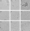
Dopa decarboxylase (Ddc) participates in melanization and nodulation. Haemocytes were incubated in Grace's culture medium in the presence of Escherichia coli and were observed under microscope (a). A developing melanization and nodule formation was obvious after 10 min (b) and 60 min (c) The presence of the Ddc inhibitors, benserazide (d) and carbidopa (e), as well as anti-Ddc (f) or anti-proPO (anti-prophenoloxidase) (g), canceled both melanization and nodule formation during the 60-min observation. A limited aggregation appeared in haemocytes treated with anti-proPO and following addition of dihydroxy-phenyl-alanine (dopa) (h) or domapine (i)

References

    1. Ashida M, Brey PT. Recent advances on the research of the insect prophenoloxidase cascade. In: Brey PT, Hultmark D, editors. Molecular Mechanisms of Immune Responses in Insects. London: Chapman & Hall; 1998. pp. 135–72.
    1. Cerenius L, Soderhall K. The prophenoloxidase-activating system in invertebrates. Immunol Rev. 2004;198:116–26. - PubMed
    1. Kanost MR, Jiang H, Yu XQ. Innate immune responses of a lepidopteran insect, Manduca sexta. Immunol Rev. 2004;198:97–105. - PubMed
    1. Mavrouli MD, Tsakas S, Theodorou GL, Lampropoulou M, Marmaras VJ. MAP kinases mediate phogocytosis and melanization via prophenoloxidase activation in medfly haemocytes. Biochim Biophys Acta. 2005;1744:145–56. - PubMed
    1. Lavine MD, Strand MR. Insect haemocytes and their role in immunity. Insect Biochem Mol Biol. 2002;32:1295–309. - PubMed

Publication types

MeSH terms

LinkOut - more resources