. 2007 Nov 13;104(46):18280-5.
doi: 10.1073/pnas.0706900104. Epub 2007 Nov 2.

Specific developmental disruption of disrupted-in-schizophrenia-1 function results in schizophrenia-related phenotypes in mice

Affiliations

Specific developmental disruption of disrupted-in-schizophrenia-1 function results in schizophrenia-related phenotypes in mice

Weidong Li et al. Proc Natl Acad Sci U S A. .

Abstract

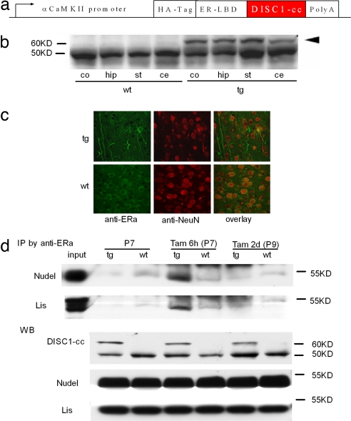
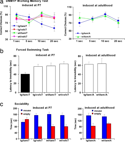
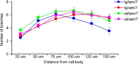
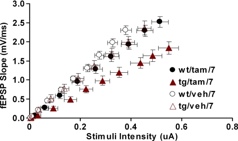
Disrupted-in-schizophrenia 1 (DISC1) was initially discovered through a balanced translocation (1;11)(q42.1;q14.3) that results in loss of the C terminus of the DISC1 protein, a region that is thought to play an important role in brain development. Here, we use an inducible and reversible transgenic system to demonstrate that early postnatal, but not adult induction, of a C-terminal portion of DISC1 in mice results in a cluster of schizophrenia-related phenotypes, including reduced hippocampal dendritic complexity, depressive-like traits, abnormal spatial working memory, and reduced sociability. Accordingly, we report that individuals in a discordant twin sample with a DISC1 haplotype, associating with schizophrenia as well as working memory impairments and reduced gray matter density, were more likely to show deficits in sociability than those without the haplotype. Our findings demonstrate that alterations in DISC1 function during brain development contribute to schizophrenia pathogenesis.


Conflict of interest statement

The authors declare no conflict of interest.

Figures

Fig. 1.
Fig. 1.
Deriving the inducible DISC1-cc transgenic mouse. (a) Schematic of the inducible DISC1-cc transgene construct. αCaMKII, α-calmodulin kinase II. (b) Western blot showing the expression of the DISC1-cc protein in the cortex (co), hippocampus (hip), striatum (st), and cerebellum (ce) of transgenic (tg) and wild-type (wt) mice. The arrowhead indicates the DISC1-cc protein, and the lower bands are the endogenous estrogen receptor. (c) Immunocytochemical labeling showing the distribution of the transgene protein in cytoplasm and dendrites of hippocampal cells. Green indicates the transgene protein or endogenous estrogen receptor, and red indicates the neuronal nucleus. (d) Immunoprecipitation assay shows that DISC1-cc protein only binds with Nudel and Lis1 6 h after tamoxifen induction and not under other conditions. The Western blot (WB) shows expression level of DISC1-cc, Nudel, and Lis1 in transgenic (tg) or wild-type (wt) mice.
Fig. 2.
Fig. 2.
Behavioral studies of the inducible DISC1-cc transgenic mouse. (a) DNMTP task. Adult DISC1-cc transgenic mice with induction at postnatal day 7 (tg/tam/7) have a significantly decreased ratio of correct DNMTP choices compared with all other groups. (b) Forced swimming test. The tg/tam/7 mice cease swimming significantly sooner than all other groups. (c) Sociability test. The tg/tam/7 mice spent equal amounts of time in the stimulus mouse chamber and the empty chamber away from the stimulus mouse. All other groups spent significantly more time in the stimulus mouse chamber than in the empty chamber.
Fig. 3.
Fig. 3.
Reduced dendritic complexity. The DiI fluorescent rapid neuronal labeling study showed that the tg/tam/7 mice had a reduced number of dendritic branches of granule cell in dentate gyrus compared with all other groups. Branching appears normal toward the soma but is deficient at distances beyond 150 μm from the soma.
Fig. 4.
Fig. 4.
Reduced hippocampal synaptic transmission in DISC1 mice. I/O relationship between the fiber volley amplitude and fEPSP slope was determined over a range of stimulus intensities (0–100 μA). Each point represents the mean of all slices tested for certain stimulus intensities. Error bars illustrate standard error for both axes. ANOVA analysis showed that the tg/tam/7 mice had reduced synaptic I/O function compared with the other three groups.

References

    1. Cannon TD, Bearden CE, Hollister JM, Rosso IM, Sanchez LE, Hadley T. Schizophr Bull. 2000;26:379–393. - PubMed
    1. Selemon LD, Goldman-Rakic PS. Biol Psychiatry. 1999;45:17–25. - PubMed
    1. Lipska BK, Weinberger DR. Neurotox Res. 2002;4:469–475. - PubMed
    1. Arnold SE, Talbot K, Hahn CG. Prog Brain Res. 2005;147:319–345. - PubMed
    1. Weinberger DR. Arch Gen Psychiatry. 1987;44:660–669. - PubMed

Publication types

LinkOut - more resources