. 2007;35(22):7698-713.
doi: 10.1093/nar/gkm538. Epub 2007 Nov 5.

Characterization of the G-quadruplexes in the duplex nuclease hypersensitive element of the PDGF-A promoter and modulation of PDGF-A promoter activity by TMPyP4

Affiliations

Characterization of the G-quadruplexes in the duplex nuclease hypersensitive element of the PDGF-A promoter and modulation of PDGF-A promoter activity by TMPyP4

Yong Qin et al. Nucleic Acids Res. 2007.

Abstract

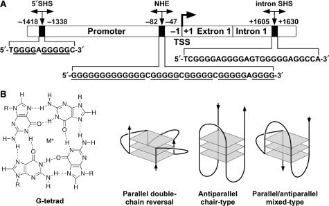
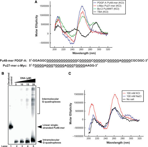
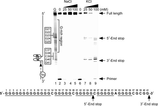
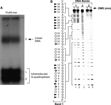
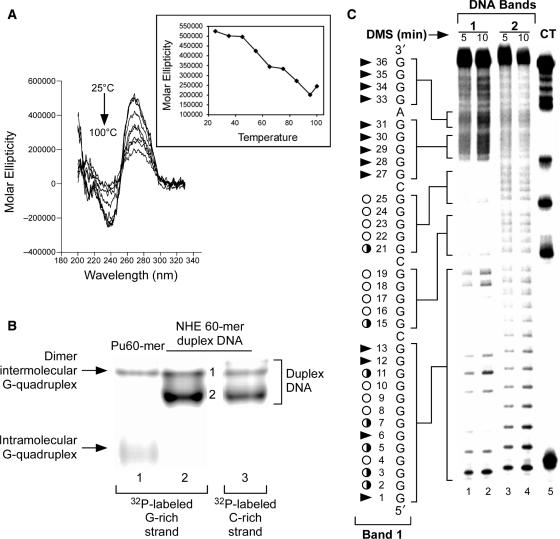
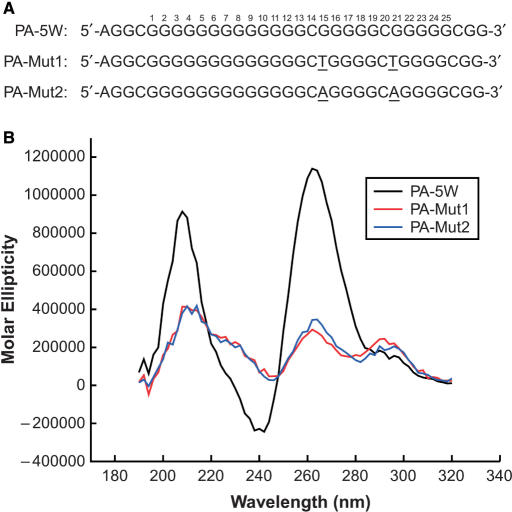
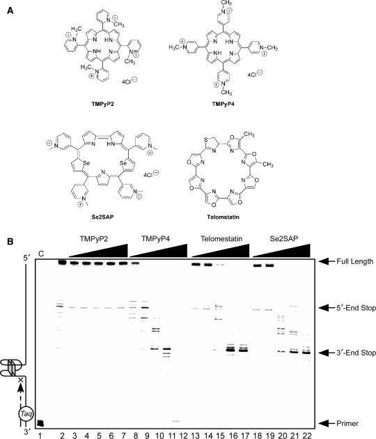
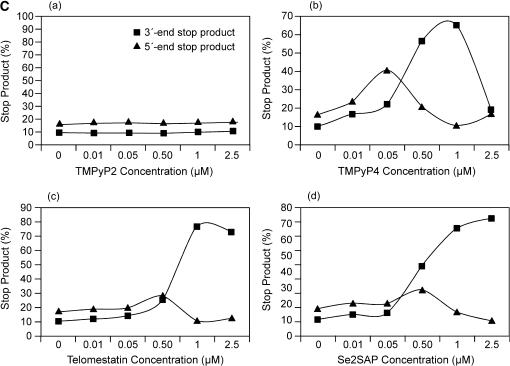
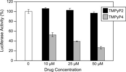
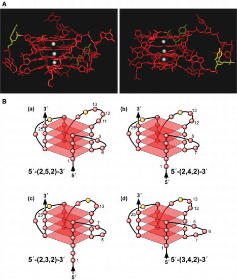
The proximal 5'-flanking region of the human platelet-derived growth factor A (PDGF-A) promoter contains one nuclease hypersensitive element (NHE) that is critical for PDGF-A gene transcription. On the basis of circular dichroism (CD) and electrophoretic mobility shift assay (EMSA), we have shown that the guanine-rich (G-rich) strand of the DNA in this region can form stable intramolecular parallel G-quadruplexes under physiological conditions. A Taq polymerase stop assay has shown that the G-rich strand of the NHE can form two major G-quadruplex structures, which are in dynamic equilibrium and differentially stabilized by three G-quadruplex-interactive drugs. One major parallel G-quadruplex structure of the G-rich strand DNA of NHE was identified by CD and dimethyl sulfate (DMS) footprinting. Surprisingly, CD spectroscopy shows a stable parallel G-quadruplex structure formed within the duplex DNA of the NHE at temperatures up to 100 degrees C. This structure has been characterized by DMS footprinting in the double-stranded DNA of the NHE. In transfection experiments, 10 microM TMPyP4 reduced the activity of the basal promoter of PDGF-A approximately 40%, relative to the control. On the basis of these results, we have established that ligand-mediated stabilization of G-quadruplex structures within the PDGF-A NHE can silence PDGF-A expression.


Figures

Figure 1.
Figure 1.
(A) Promoter structure of PDGF-A and locations of the important NHEs. The sequences of the 5′SHS, the G-rich strand of the NHE and the intron SHS are shown. TSS = transcriptional start site. (B) Structure of a G-tetrad and examples of the folding patterns of known intramolecular G-quadruplexes.
Figure 2.
Figure 2.
(A) Comparative CD spectra of three known G-quadruplex-forming sequences with the PDGF-A Pu48-mer. Blue line = PDGF-A Pu48-mer (25 mM KCl), red line = c-Myc Pu27-mer (parallel G-quadruplex in 25 mM KCl), green line = Bcl-2 Pu39WT (mixed parallel/antiparallel G-quadruplex in 100 mM KCl), black line = TBA (antiparallel G-quadruplex in 100 mM KCl). All CD data were obtained with a 5 μM strand concentration at 25°C. Comparison of sequences of these two DNA oligomers is shown under the CD spectra. (B) Native gel electrophoresis indicating the formation of intramolecular G-quadruplex structures by Pu48-mer. The fast mobility major band represents the intramolecular G-quadruplex and the slow mobility bands represent the higher-order intermolecular G-quadruplexes. (C) Effect of alkali metals on the Pu48-mer CD spectra. Effect of KCl (red line) and NaCl (blue line) on the ellipticity signal compared to the signal in the absence of salt (black line). All CD data were obtained with a 5 μM strand concentration at 25°C.
Figure 3.
Figure 3.
Effect of K+ and Na+ on the formation of the NHEPDGF-A G-quadruplex in a Taq polymerase stop assay. Two stop products are designated as the 5′-end product and the 3′-end stop product. The corresponding arrest sites are indicated on the core G-tract sequence.
Figure 4.
Figure 4.
The EMSA (A) and DMS footprinting (B) of intramolecular G-quadruplex structures of PDGF-A Pu48-mer. In (A), high-mobility bands 1 and 2 represent compact intramolecular G-quadruplex structures, and band 3 represents linear DNA, which was fully denatured by heating. In (B), DMS footprinting of band 1 (lanes 1 and 2), band 2 (lanes 4 and 5) and band 3 (lane 3) from (A) is shown. Open circles indicate the guanines that are fully protected, partially open circles indicate the guanines that are partially protected and arrowheads indicate the guanines that are cleaved.
Figure 5.
Figure 5.
(A) Effect of temperature (25–100°C) on the CD spectra of 90 bp duplex DNA containing NHEPDGF-A. At 100°C, 90 bp duplex DNA of NHEPDGF-A still generated a strong G-quadruplex CD signal. (B) Non-denatured gel analysis of 60-mer G-rich single-stranded DNA of NHEPDGF-A (lane 1) and 60 bp double-stranded DNA of NHEPDGF-A (lanes 2 and 3). In lanes 1 and 2, the G-rich strand was 5′-end-radiolabeled with 32P, and the C-rich strand was 5′-end-radiolabeled with 32P in lane 3. (C) DMS footprinting for G-rich strands of 60 bp duplex DNA band 1 (lanes 1 and 2) and band 2 (lanes 3 and 4). Lane 5 shows the CT sequencing on the G-rich strand of the 60 bp duplex DNA of NHEPDGF-A. Open circles indicate the guanines that are fully protected, partially open circles indicate the guanines that are partially protected, and arrowheads indicate the guanines that are cleaved.
Figure 6.
Figure 6.
Sequences (A) and comparative CD spectra (B) of the wild-type sequence of the three 5′-end runs of guanines and the G15/G21 double mutation sequences in the presence of 100 mM KCl. Black line = wild-type sequence PA-5W, red line = double G-to-T mutation sequence PA-Mut1 and blue line = double G-to-A mutation sequence PA-Mut2. All CD data were obtained with a 5 μM strand concentration at 25°C.
Figure 7.
Figure 7.
(A) Structures of the G-quadruplex-interactive compounds TMPyP4, telomestatin and Se2SAP, and the control compound TMPyP2. (B) The Taq polymerase stop assay was used to compare the stabilization of the NHEPDGF-A G-quadruplex by TMPyP2 (lanes 3–7), TMPyP4 (lanes 8–12), telomestatin (lanes 13–17) and Se2SAP (lanes 18–22) by using increasing concentrations of drugs (0.01, 0.05, 0.5, 1 and 2.5 μM) at 60°C. Lane 1 is control, and lane 2 is without drug. (C) The ratios of the major arrest products of each sample to the total product were plotted against drug concentrations.
Figure 7.
Figure 7.
(A) Structures of the G-quadruplex-interactive compounds TMPyP4, telomestatin and Se2SAP, and the control compound TMPyP2. (B) The Taq polymerase stop assay was used to compare the stabilization of the NHEPDGF-A G-quadruplex by TMPyP2 (lanes 3–7), TMPyP4 (lanes 8–12), telomestatin (lanes 13–17) and Se2SAP (lanes 18–22) by using increasing concentrations of drugs (0.01, 0.05, 0.5, 1 and 2.5 μM) at 60°C. Lane 1 is control, and lane 2 is without drug. (C) The ratios of the major arrest products of each sample to the total product were plotted against drug concentrations.
Figure 8.
Figure 8.
Dual luciferase assay to determine the effect of TMPyP4 and TMPyP2 on the transcriptional activity of PDGF-A basal promoter containing the NHE. The comparative firefly luciferase expressions (firefly/renilla) of TMPyP2 and TMPyP4 are shown in the histograms. The values are the average of three independent experiments. Error bars are ± SE.
Figure 9.
Figure 9.
(A) Model of the biologically relevant NHEPDGF-A G-quadruplex [loop isomer 5′-(2,5,2)-3′], which contains two 2-base double-chain reversal loops and one 5-base intervening loop (guanines = red, cytosines = yellow and K+ ions = white). For clarity, hydrogen atoms have been removed. In the left panel, the two 2-base double-chain reversal loops are shown on each side of model, and in the right panel, the model has been rotated to show the 5-base intervening loop on the right side of model. (B) Proposed folding patterns of the four different loop isomers formed in the core sequence of NHEPDGF-A. Guanines = red, cytosines = yellow.

References

    1. Heldin CH, Westermark B. Mechanism of action and in vivo role of platelet-derived growth factor. Physiol. Rev. 1999;79:1283–1316. - PubMed
    1. Heldin CH, Eriksson U, Ostman A. New members of the platelet-derived growth factor family of mitogens. Arch. Biochem. Biophys. 2002;398:284–290. - PubMed
    1. Alvarez RH, Kantarjian HM, Cortes JE. Biology of platelet-derived growth factor and its involvement in disease. Mayo Clin. Proc. 2006;81:1241–1257. - PubMed
    1. Betsholtz C. Biology of platelet-derived growth factors in development. Birth Defects Res. C Embryo. Today. 2003;69:272–285. - PubMed
    1. Ostman A, Heldin CH. Involvement of platelet-derived growth factor in disease: development of specific antagonists. Adv. Cancer Res. 2001;80:1–38. - PubMed

Publication types