. 2007;35(22):7688-97.
doi: 10.1093/nar/gkm912. Epub 2007 Nov 4.

Saccharomyces cerevisiae Ebs1p is a putative ortholog of human Smg7 and promotes nonsense-mediated mRNA decay

Affiliations

Saccharomyces cerevisiae Ebs1p is a putative ortholog of human Smg7 and promotes nonsense-mediated mRNA decay

Brian Luke et al. Nucleic Acids Res. 2007.

Abstract

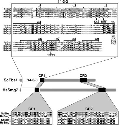
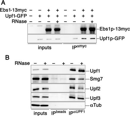
The Smg proteins Smg5, Smg6 and Smg7 are involved in nonsense-mediated RNA decay (NMD) in metazoans, but no orthologs have been found in the budding yeast Saccharomyces cerevisiae. Sequence alignments reveal that yeast Ebs1p is similar in structure to the human Smg5-7, with highest homology to Smg7. We demonstrate here that Ebs1p is involved in NMD and behaves similarly to human Smg proteins. Indeed, both loss and overexpression of Ebs1p results in stabilization of NMD targets. However, Ebs1-loss in yeast or Smg7-depletion in human cells only partially disrupts NMD and in the latter, Smg7-depletion is partially compensated for by Smg6. Ebs1p physically interacts with the NMD helicase Upf1p and overexpressed Ebs1p leads to recruitment of Upf1p into cytoplasmic P-bodies. Furthermore, Ebs1p localizes to P-bodies upon glucose starvation along with Upf1p. Overall our findings suggest that NMD is more conserved in evolution than previously thought, and that at least one of the Smg5-7 proteins is conserved in budding yeast.


Figures

Figure 1.
Figure 1.
The Smg5-7 14-3-3 phosphoserine-binding residues are conserved in Ebs1p. Multiple sequence alignment of the 14-3-3 domains of human Smg5 (amino acids 19–266), Smg6 (576–818) and Smg7 (1–236) proteins with the N-terminal regions of yeast Ebs1 (23–291) and Est1 (22–291) proteins. The alignment was produced using the ClustalW program. Alpha helices 1–9 (α1–α 9) are shown. Conserved amino acids are indicated as white letters on a black background while similar amino acids are white letters on a gray background. Arrowheads point to consensus 14-3-3 residues (indicated below each arrowhead) derived from the human 14-3-3 zeta isoform that have been implicated in phosphoserine binding for 14-3-3 domains across species. Black arrowheads indicate 14-3-3 residues that are conserved with respect to side-chain charge content in Ebs1p but not in Est1p, the white arrowhead points to a residue conserved in both Ebs1p and Est1p, the gray arrowhead highlights a residue, which is not conserved in either of the two yeast proteins. The cartoon depicts the domain architecture of yeast Ebs1 and human Smg7 proteins. Two conserved regions named CR1 and CR2 are indicated as black boxes and their sequence is shown in the lower insets. CR1: amino acids 231–283 in Ebs1p and 174–226 in Smg7; CR2: amino acids 710–789 in Ebs1p and 933–1013 in Smg7.
Figure 2.
Figure 2.
Ebs1 participates in NMD in a way similar to human Smg proteins. (A and B) Total RNA from log phase yeast grown in YPD was prepared from wt, upf1Δ, ebs1Δ, est1Δ, ebs1Δest1Δ and ebs1Δupf1Δ cells and subjected to northern blot analysis using a probe which detects both the CYH2 pre-mRNA and CYH2 mRNA. In (A) the filter was stripped and re-probed to detect the ade2-1 mRNA. CYH2 pre-mRNA levels (CYH2 ratio) and ade2-1 mRNA levels (ade2-1 ratio) are expressed relative to wild-type cells, after normalization using the CYH2 mRNA signal. (C) HeLaβGWT and HeLaβGNS39 cells (see Materials and Methods section) were transfected with shRNA vectors against human Smg7 (sh7A and sh7B), human Smg6 (sh6) and human Upf1 and with empty vector controls (shEV). Five days after transfection, total mRNA was prepared and the levels of β-globin mRNA were determined on a northern blot. Alternatively, the same cell lines were transfected with vectors expressing a Flag-tagged version of human Smg7 (F-Smg7) or empty vector controls (F-EV) and processed 48 h after transfection. The signal of βGNS39 (βGL ratio) is expressed relative to empty vector-transfected cells, after normalization using the actin signal (loading control) and the signal of βGWT. (D) Wild-type yeasts were transformed with centromeric plasmids harboring EBS1 and Smg7 under the constitutive ADH promoter, or with empty vector alone. Total RNA was probed using the CYH2 probe as in A–C.
Figure 3.
Figure 3.
Ebs1p and Upf1p co-localize in P-bodies in glucose-starved cells. Yeast cells expressing Ebs1p-dsRED and Dcp2p-GFP fusions as well as cells expressing Ebs1p-dsRED and Upf1p-GFP were grown in rich synthetic media and successively resuspended in media lacking glucose while shaking at 30°C. Four hours after, confocal images of live cells were taken. Arrows point to cytoplasmic foci where a co-localization between Ebs1p-dsRED with Dcp2p-GFP and Ebs1p-dsRED with Upf1p-GFP occurred. Scale bar represents 2.5 μm.
Figure 4.
Figure 4.
Overexpression of Ebs1 recruits Upf1p into P-bodies. Upf1p-GFP and Dcp2p-RFP cells were transformed with either empty vector (EV) or with a plasmid, which overexpressed Ebs1p under the ADH promoter (ADH-Ebs1) and subjected to confocal microscopy. Arrowheads indicate examples of cytoplasmic foci where a co-localization between Upf1p-GFP and Dcp2p-RFP occurred. At the time of imaging, 74% of the cells transformed with ADH-Ebs1 displayed 2–6 Upf1p-GFP foci and 6% of the cells transformed with empty vectors displayed 1 to 2 Upf1p-GFP foci. Scale bar represents 2.5 μm.
Figure 5.
Figure 5.
Smg7/Ebs1p physically interacts with Upf1 proteins in a partially RNA-dependent manner. (A) Myc tagged Ebs1p was immunoprecipitated from cell lysates prepared from strains expressing Upf1p-GFP. Lysates were treated with RNase or left untreated. Western blots were performed using anti-myc and anti-GFP antibodies. (B) Protein extracts were prepared form HeLa cells and treated with RNase or left untreated. Human Upf1 was immunoprecipitated using goat polyclonal antibodies and western blot analysis was performed using rabbit polyclonal antibodies raised against human Smg7, Upf2 and Upf3 and goat polyclonal antibodies raised against Upf1. As a control for IP specificity, α-Tubulin does not co-immunoprecipitate with Upf1.

References

    1. Maquat LE. Nonsense-mediated mRNA decay: splicing, translation and mRNP dynamics. Nat. Rev. Mol. Cell. Biol. 2004;5:89–99. - PubMed
    1. Wittmann J, Hol EM, Jack HM. hUPF2 silencing identifies physiologic substrates of mammalian nonsense-mediated mRNA decay. Mol. Cell. Biol. 2006;26:1272–1287. - PMC - PubMed
    1. Azzalin CM, Lingner J. The human RNA surveillance factor UPF1 is required for S phase progression and genome stability. Curr. Biol. 2006;16:433–439. - PubMed
    1. Anders KR, Grimson A, Anderson P. SMG-5, required for C.elegans nonsense-mediated mRNA decay, associates with SMG-2 and protein phosphatase 2A. EMBO J. 2003;22:641–650. - PMC - PubMed
    1. Leeds P, Peltz SW, Jacobson A, Culbertson MR. The product of the yeast UPF1 gene is required for rapid turnover of mRNAs containing a premature translational termination codon. Genes Dev. 1991;5:2303–2314. - PubMed

Publication types

MeSH terms