. 2008 Jan 4;283(1):603-612.
doi: 10.1074/jbc.M708250200. Epub 2007 Nov 5.

Rho GTPase and cAMP/protein kinase A signaling mediates myocilin-induced alterations in cultured human trabecular meshwork cells

Affiliations

Rho GTPase and cAMP/protein kinase A signaling mediates myocilin-induced alterations in cultured human trabecular meshwork cells

Xiang Shen et al. J Biol Chem. .

Abstract

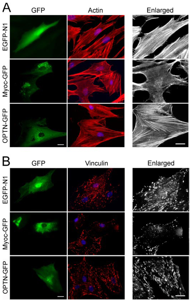
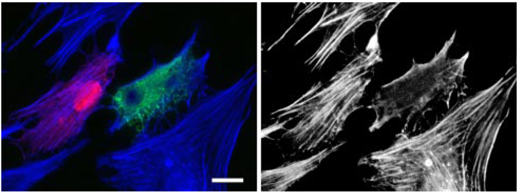
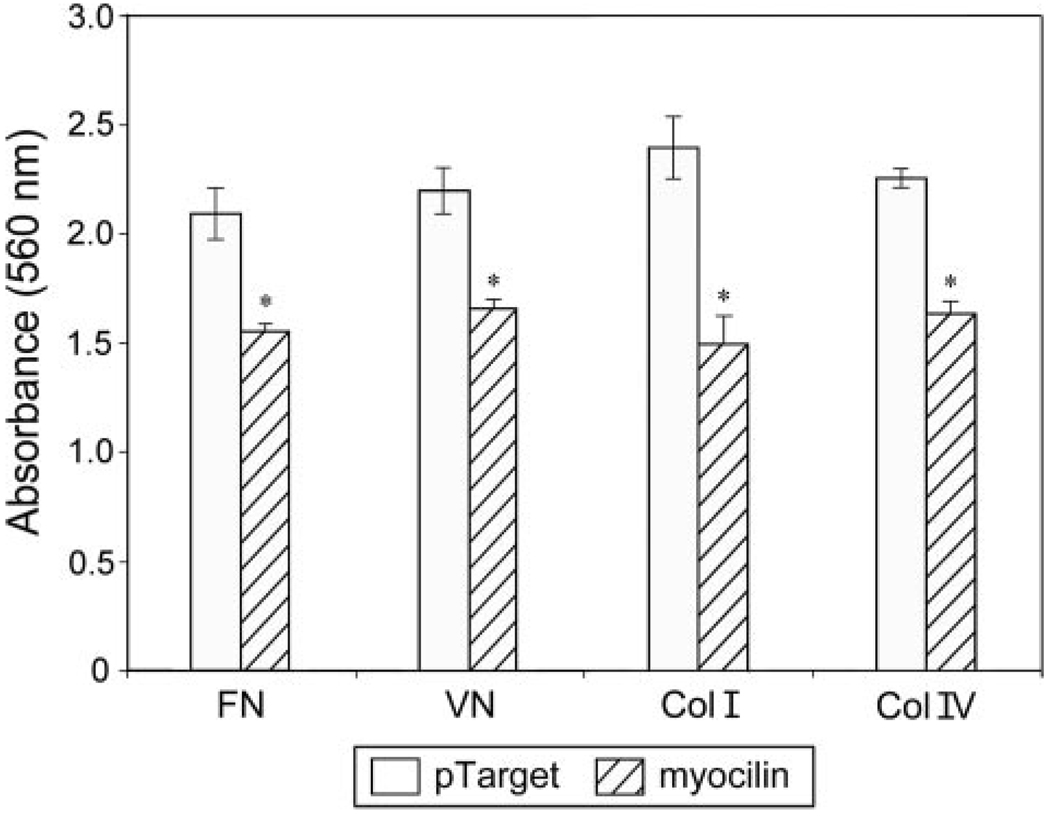
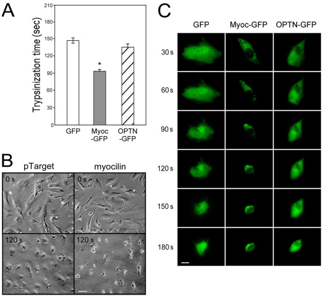
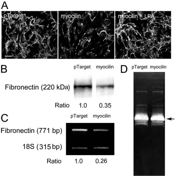
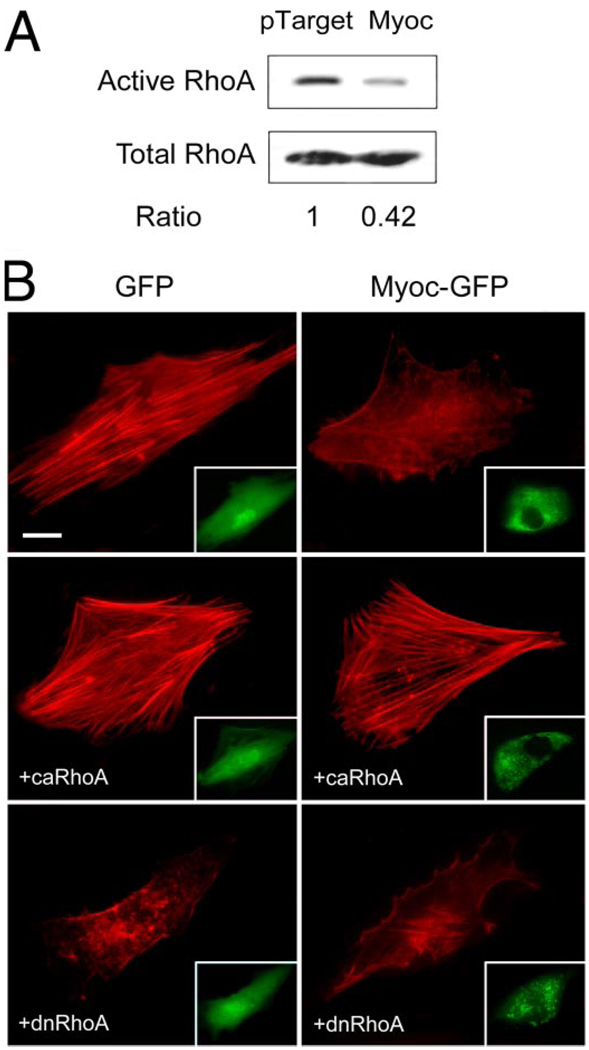
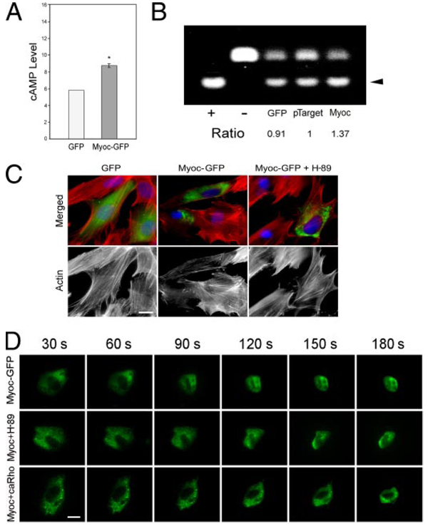
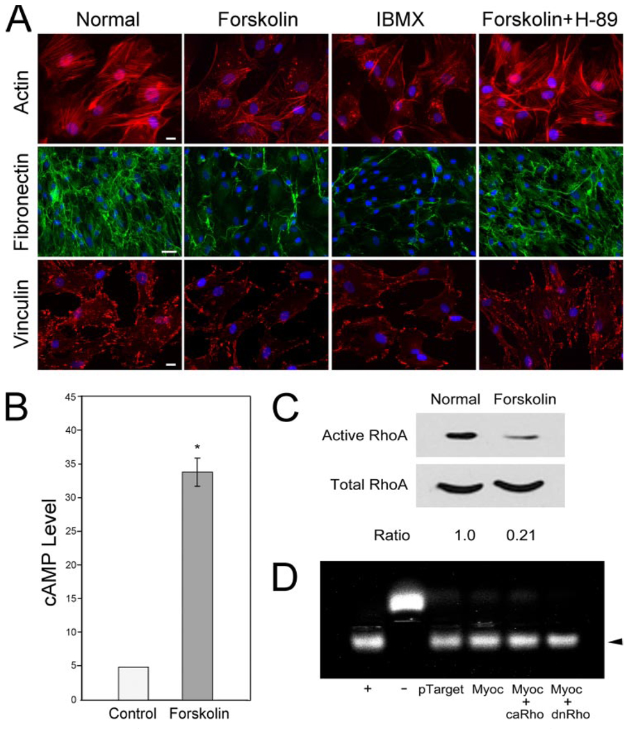
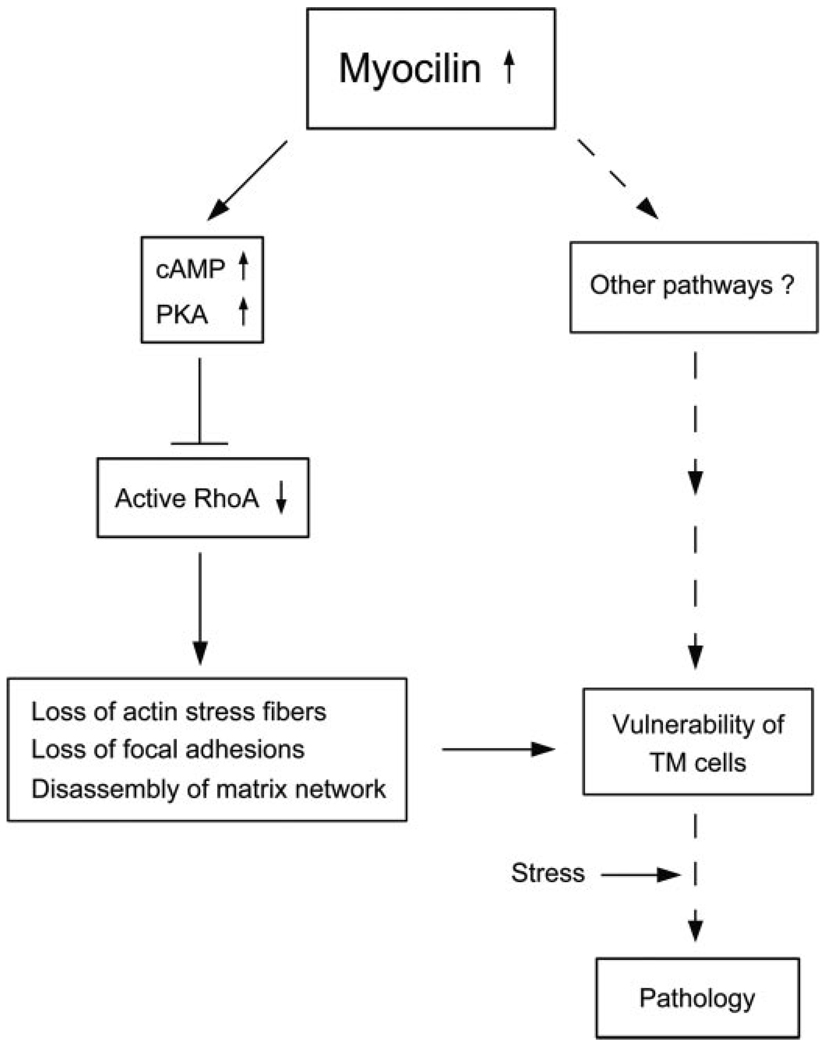
Myocilin is a gene linked to the most common form of glaucoma, a major blinding disease. The trabecular meshwork (TM), a specialized eye tissue, is believed to be involved, at least in part, in the development of glaucoma. The myocilin expression is known to be up-regulated by glucocorticoids in TM cells, and an altered myocilin level may be the culprit in conditions such as corticosteroid glaucoma. Wild type myocilin, when transfected into cultured human TM cells, induced a dramatic loss of actin stress fibers and focal adhesions. Myocilin transfectants displayed a heightened sensitivity to trypsin. Adhesion to fibronectin, collagens, and vitronectin was compromised. The fibronectin deposition and the levels of fibronectin protein and mRNA were also reduced in myocilin transfectants. The fibronectin deposition could be restored by treatment with lysophosphatidic acid, a Rho stimulator. Assays further revealed that upon myocilin overexpression, the activity of RhoA was diminished, whereas the cAMP level and the protein kinase A (PKA) activity were augmented. Myocilin protein did not affect actin polymerization. The collapse of actin stress fibers and increased trypsin sensitivity from myocilin transfection could be reverted by co-expression of constitutively active RhoA or by treatment with PKA inhibitor H-89. The PKA activity, however, was not modified by co-expression of either constitutively active or dominant negative RhoA. These results demonstrate that myocilin has a de-adhesive activity and triggers signaling events. cAMP/PKA activation and the downstream Rho inhibition are possible mechanisms by which myocilin in overabundance may lead to TM cell or tissue damage.


Figures

FIGURE 1
FIGURE 1. Myocilin overexpression induces a loss of actin stress fibers (A) and vinculin (B) staining
TM cells were transfected with pEGFP-N1 (EGFP-N1; mock control), pMyocilin-EGFP (Myoc-GFP), or pOptineurin-EGFP (OPTN-GFP; a negative control) and stained for actin (A, red) or vinculin (B, red). The transfected cells were marked by green fluorescence. The enlarged actin and vinculin micrographs of the transfected cells are presented in black and white. The staining was visualized using a Zeiss 100M microscope. Bar, 20 µm.
FIGURE 2
FIGURE 2. Actin staining in mixed cultures of pMyocilin-EGFP- and pDsRed-Monomer-C1-transfected cells
F-actin was stained with Alexa Fluor 350-phalloidin in blue. The actin cytoskeleton is shown in black and white in the right panel. The pMyocilin-EGFP-transfected cell (in green) showed a loss of actin stress fibers, whereas the DsRed-expressing (in red) and nontransfected cells had robust actin fibers. Bar, 20 µm.
FIGURE 3
FIGURE 3. Myocilin overexpression impairs TM cell adhesion
Adhesion of pTarget (mock control)- and pTarget-myocilin (myocilin)-transfected TM cells was determined using Chemicon CytoMatrix cell adhesion strips coated with fibronectin, vitronectin, and collagen types I and IV. Data are presented as mean ± S.E. (n = 3). Asterisks, data significantly different from corresponding pTarget mock controls. The p values for fibronectin, vitronectin, and collagen type I and IV were 0.0046, 0.0032, 0.0063, and < 0.0001, respectively.
FIGURE 4
FIGURE 4. Myocilin-transfected cells displayed a heightened sensitivity to trypsinization
A, the trypsinization time needed for pEGFP-N1 (GFP)-, pMyocilin-EGFP (Myoc-GFP)-, and pOptineurin-EGFP (OPTN-GFP)- transfected TM cells to become refractile. *, the trypsinization time for myocilin transfectants was significantly (p < 0.0001, n = 30) lower than that of GFP or optineurin controls. B, phase-contrast micrographs of pTarget- and pTarget-myocilin (myocilin)-transfected cells before (0 s) or after trypsinization for 120 s (120 s). Scale bar, 50 µm. C, time lapse video microscopy of pEGFP-N1 (GFP)-, pMyocilin-EGFP (Myoc-EGFP)-, and pOptineurin-EGFP (OPTN-GFP)-transfected cells every 30 s after the addition of trypsin solution to the cultures. Bar, 20 µm.
FIGURE 5
FIGURE 5
A, effect of myocilin transfection and/or LPA on fibronectin assembly in human TM cultures. Fibronectin (in green) assembly in pTarget-transfected, pTarget-myocilin (myocilin)-transfected, and myocilin-transfected and LPA (5 µM, 16 h)-treated (myocilin + LPA) TM cells was visualized by immunofluorescence. The nuclei were stained by 4′,6′-diamidino- 2-phenylindole dihydrochloride in blue. Experiments were repeated three times, yielding similar results. Bar, 50 µm. B, secreted fibronectin protein level; C, fibronectin transcript level; D,MMPactivities in pTarget- and pTarget-myocilin (myocilin)-transfected TM cultures. Fibronectin secretion into the medium was examined by Western blotting (B). The level of fibronectin transcript was measured by relative quantitative RT-PCR (C). Relative to mock controls, the secreted fibronectin protein and the transcript levels were reduced by ~70%. The MMP activities were visualized by zymography (D). Experiments were repeated two times, yielding similar results.
FIGURE 6
FIGURE 6. Effect of recombinant human myocilin on actin polymerization
In in vitro assays, the actin polymerization was induced by actin polymerization buffer in the absence or presence of 90 nm, 900 nm, or 9µm recombinant human myocilin. The actin polymerization buffer was not added to the negative control. Experiments were repeated three times with similar results.
FIGURE 7
FIGURE 7
A, GTP-bound active RhoA in pTarget- and pTarget-myocilin (Myoc)-transfected TM cells. Pull-down assays were performed to determine the RhoA activity. The amount of the active or GTP-bound RhoA was normalized against the total amount in cell lysates. Data compiled indicated that the RhoA activity upon myocilin transfection (0.55 ± 0.10, n = 6) was significantly reduced (p < 0.007) compared with controls. B, effect of co-expression of ca or dn RhoA in TM cells on the actin cytoskeleton. Human TM cells in culture were transfected with pEGFP-N1 (GFP) or pMyocilin-EGFP (Myoc-GFP) with-out (top panels), or with co-transfection with plasmid encoding ca V14 RhoA (+caRhoA) or dn N19 RhoA (+dnRhoA). The transfected cells displaying green fluorescence are shown in the insets. The actin staining (in red) was examined by fluorescence microscopy. Cells transfected with pEGFP-N1 as a control exhibited robust actin stress fibers. Transfection with pMyocilin-EGFP caused a loss of actin fibers. An increase of the actin assembly or rescue of the myocilin phenotype was observed with co-expression of the ca but not dn form of RhoA. Bar, 20 µm.
FIGURE 8
FIGURE 8
A, cAMP level in pEGFP-N1 (GFP)- or pMyocilin-EGFP (Myoc-GFP)- transfected TM cells. The cAMP level in myocilin transfectants (1.47 ± 0.03, n = 4) measured by an ELISA was expressed relative to the controls. *, the cAMP level for myocilin transfectants was significantly (p < 0.0037) increased compared with GFP mock controls. B, PKA activity in pEGFP-N1 (GFP)-, pTarget-, or pTarget-myocilin (Myoc)-transfected cells. Equal amounts of protein lysates were subjected to PKA assays. Positive (+) and negative (−) controls were included. The nonphosphorylated (upper band) and the phosphorylated (lower band, arrowhead) substrates were resolved on agarose gels. The PKA activity, judged by the level of the phosphorylated substrate, in pTarget-myocilin transfectants (1.39 ± 0.02, n = 9, p < 0.0001) was determined by densitometric analyses and normalized to that in pTarget controls. Results shown are from one of the experiments. C, actin staining in pEGF-N1 (GFP)- and pMyocilin-EGFP (Myoc-GFP)-transfected TM cells without or with the treatment with PKA inhibitor, H-89 (Myoc + H-89). D, time lapse video microscopy of pMyocilin-EGFP (Myoc-GFP)-transfected cells and those with H-89 treatment (Myoc + H-89) or co-transfection with ca RhoA (Myoc + caRho) every 30 s after the addition of trypsin solution to the cultures. Bar, 20 µm.
FIGURE 9
FIGURE 9
A, actin (red), fibronectin (green), and vinculin (red) staining in normal humanTMcells after overnight treatment with 50 µm forskolin, 500 µm IBMX, or 50 µm forskolin plus 10 nM H-89. The nuclei were stained by 4′,6′-diamidino-2-phenylindole dihydrochloride in blue. Bar for actin and vinculin, 20 µm; bar for fibronectin, 50 µm. B, cAMP level was increased by forskolin treatment by ~7-fold (34.0 ± 6.7 versus 4.9 ± 0.1; n = 4). C, inhibition of RhoA activity after forskolin induction ofcAMPin normalhumanTMcells. The experiments were repeated twice. D, PKA activity in TM cells transfected with pTarget, pTarget-myocilin (Myoc), pTarget-myocilin plus ca RhoA (Myoc + caRho), or pTarget-myocilin plus dn RhoA (Myoc + dnRho). Positive (+) and negative (−) controls were included. The PKA activity was determined as described in the legend to Fig. 8B. Results were expressed as ratios relative to the pTarget control. Data from one representative experiment are presented.
FIGURE 10
FIGURE 10. A working model of possible events in human TM cells triggered by up-regulation of myocilin
Myocilin, when moderately up-regulated, induces cAMP/PKA activation and the downstream RhoA inactivation, leading to a loss of actin stress fibers and focal adhesions and disassembly of matrix network. These changes or other pathways may affect the integrity of TM cells, rendering them more susceptible to additional stress or challenge and pathologic consequences. Other intermediate mediators or pathways have yet to be identified. The dotted lines indicate steps that have not been investigated in the present work or in our previous investigation (17).

References

    1. Bill A. Invest. Ophthalmol. Vis. Sci. 1975;14:1–3. - PubMed
    1. Yue BYJT. Surv. Ophthalmol. 1996;40:379–390. - PubMed
    1. Zhou L, Fukuchi T, Kawa JE, Higginbotham EJ, Yue BYJT. Invest. Ophthalmol. Vis. Res. 1995;36:787–795. - PubMed
    1. Calthorpe CM, Grierson I. Exp. Eye Res. 1990;51:39–48. - PubMed
    1. Stumpff F, Wiederholt M. Ophthalmologica. 2000;214:33–53. - PubMed

Publication types

MeSH terms