. 2008 Jan;76(1):361-8.
doi: 10.1128/IAI.01199-07. Epub 2007 Nov 5.

Enteropathogenic Escherichia coli O125:H6 triggers attaching and effacing lesions on human intestinal biopsy specimens independently of Nck and TccP/TccP2

Affiliations

Enteropathogenic Escherichia coli O125:H6 triggers attaching and effacing lesions on human intestinal biopsy specimens independently of Nck and TccP/TccP2

Li Bai et al. Infect Immun. 2008 Jan.

Abstract

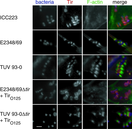
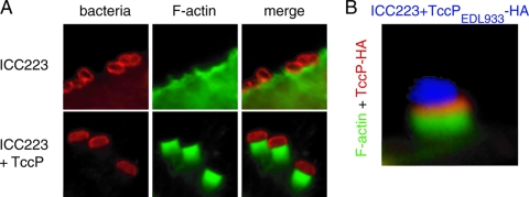
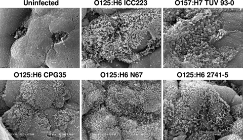
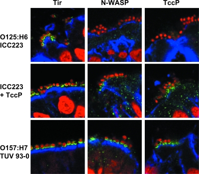
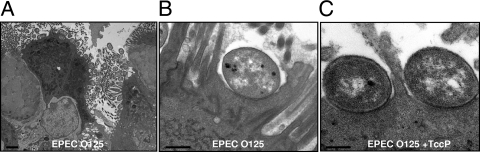
Typical enteropathogenic Escherichia coli (EPEC) and enterohemorrhagic E. coli (EHEC) employ either Nck, TccP/TccP2, or Nck and TccP/TccP2 pathways to activate the neuronal Wiskott-Aldrich syndrome protein (N-WASP) and to trigger actin polymerization in cultured cells. This phenotype is used as a marker for the pathogenic potential of EPEC and EHEC strains. In this paper we report that EPEC O125:H6, which represents a large category of strains, lacks the ability to utilize either Nck or TccP/TccP2 and hence triggers actin polymerization in vitro only inefficiently. However, we show that infection of human intestinal biopsies with EPEC O125:H6 results in formation of typical attaching and effacing lesions. Expression of TccP in EPEC O125:H6, which harbors an EHEC O157-like Tir, resulted in efficient actin polymerization in vitro and enhanced colonization of human intestinal in vitro organ cultures with detectable N-WASP and electron-dense material at the site of bacterial adhesion. These results show the existence of a natural category of EPEC that colonizes the gut mucosa using Nck- and TccP-independent mechanisms. Importantly, the results highlight yet again the fact that conclusions made on the basis of in vitro cell culture models cannot be extrapolated wholesale to infection of mucosal surfaces and that the ability to induce actin polymerization on cultured cells should not be used as a definitive marker for EPEC and EHEC virulence.


Figures

FIG. 1.
FIG. 1.
EPEC O125:H6 strain ICC223 cannot efficiently induce actin polymerization in infected HeLa cells, while the controls EPEC O127:H6 strain E2348/69 and EHEC O157:H7 strain TUV 93-0 trigger efficient actin polymerization. Expression of TirEPEC_O125:H6 in EPEC E2348/69Δtir did not restore actin polymerization, while expressing TirEPEC_O125:H6 in EHEC TUV 93-0Δtir resulted in strong actin polymerization. Bacterial DNA was visualized in blue using Hoechst 33342. Tir is labeled red with anti-TirEHEC or TirEPEC (for strain E2348/69) antiserum. Actin was labeled in green using Oregon Green-conjugated phalloidin. Separate monochrome images of the UV, red, and green fluorescence channels and a merged color image are shown. Bar, 5 μm.
FIG. 2.
FIG. 2.
A. The phylogenetic relationship of the tir genes from EPEC O125:H6 with selected previously published tir genes. B. The amino acid sequence of Tir proteins of EPEC O125:H6 strain ICC223 was aligned with previously published Tir sequences as described elsewhere (30). The tir genes of amino acid residues identical in all the proteins are indicated in black, and the residues shared by no less than 50% identity within all proteins are gray. The intimin-binding domain and two predicted transmembrane domains are indicated by a dashed line and underlining, respectively. Black triangles indicate the tyrosine residues phosphorylated by a host cell kinase(s). Underlining with *1 indicates the regions containing Y454 that are involved in pedestal formation via the TirEHEC_O157:H7-TccP/EspFu pathway and the alternative TirEPEC_O127:H6-Nck-independent pathway. Underlining with *2 indicates the TirEPEC_O127:H6 Y474 involved in the Nck pedestal formation pathway. The underlining with *3 indicates the region corresponding to the O157 EHEC Tir residues 519 to 524 that may be related to the type III secretion system-dependent secretion efficiency.
FIG. 3.
FIG. 3.
A. ICC223 binds to HeLa cells but cannot trigger efficient actin polymerization. Expression of TccPEHEC_O157:H7 confers strong actin polymerization activity. B. HA staining shows that TccP is concentrated under attached ICC223 bacteria.
FIG. 4.
FIG. 4.
Scanning electron micrographs of EPEC O125:H6 strains (ICC223, 35, N67, and 2741-5) and EHEC O157:H7 (TUV 93-0) on human terminal ileum after 8 h of IVOC. All strains show intimate adherence to the mucosa, and microvillous elongation between attaching bacteria is evident. An uninfected sample was included as a negative control. Bar, 5 μm.
FIG. 5.
FIG. 5.
Immunofluorescence staining of cryosections of human terminal ileum infected with EPEC O125:H6 (ICC223), its TccP-expressing derivative (ICC223 + TccP), and EHEC O157:H7 (TUV 93-0). Whereas all strains show Tir translocation (green) into the host cell membrane, N-WASP staining (green) can be observed underneath TccP-expressing TUV 93-0 and ICC223 + TccP but is only very weakly recruited beneath a minority of ICC223 bacteria. Sections were counterstained with propidium iodide (red) and anticytokeratin (blue) to visualize bacteria/cell nuclei and epithelial cells, respectively. Shown are merged images of all fluorescence channels.
FIG. 6.
FIG. 6.
Transmission electron microscopy of human IVOC infected with ICC223. A. ICC223 efficiently colonizes the gut mucosa. B. Typical A/E lesion with intimate bacterial attachment and effacement of brush border microvilli; increased electron density at the site of bacterial attachment (representing accumulated actin) is not apparent. C. ICC223 expressing a TccP A/E lesion, showing increased electron density in the epithelium at the site of attachment. Bars, 2 μm (A) or 0.5 μm (B and C).

References

    1. Brady, M. J., K. G. Campellone, M. Ghildiyal, and J. M. Leong. 23 May 2007. Enterohaemorrhagic and enteropathogenic Escherichia coli Tir proteins trigger a common Nck-independent actin assembly pathway. Cell. Microbiol. doi:10.1111/j.1462-5822.2007.00954.x. - DOI - PubMed
    1. Campellone, K. G., A. Giese, D. J. Tipper, and J. M. Leong. 2002. A tyrosine-phosphorylated 12-amino-acid sequence of enteropathogenic Escherichia coli Tir binds the host adaptor protein Nck and is required for Nck localization to actin pedestals. Mol. Microbiol. 431227-1241. - PubMed
    1. Campellone, K. G., and J. M. Leong. 2005. Nck-independent actin assembly is mediated by two phosphorylated tyrosines within enteropathogenic Escherichia coli Tir. Mol. Microbiol. 56416-432. - PubMed
    1. Campellone, K. G., S. Rankin, T. Pawson, M. W. Kirschner, D. J. Tipper, and J. M. Leong. 2004. Clustering of Nck by a 12-residue Tir phosphopeptide is sufficient to trigger localized actin assembly. J. Cell Biol. 164406-416. - PMC - PubMed
    1. Campellone, K. G., D. Robbins, and J. M. Leong. 2004. EspFU is a translocated EHEC effector that interacts with Tir and N-WASP and promotes Nck-independent actin assembly. Dev. Cell 7217-228. - PubMed

Publication types

MeSH terms

Associated data

LinkOut - more resources