. 2008 Jan;28(1):315-25.
doi: 10.1128/MCB.01168-07. Epub 2007 Nov 5.

Identification and analysis of a conserved Tcfap2a intronic enhancer element required for expression in facial and limb bud mesenchyme

Affiliations

Identification and analysis of a conserved Tcfap2a intronic enhancer element required for expression in facial and limb bud mesenchyme

Weiguo Feng et al. Mol Cell Biol. 2008 Jan.

Abstract

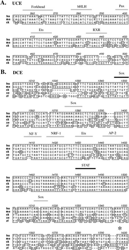
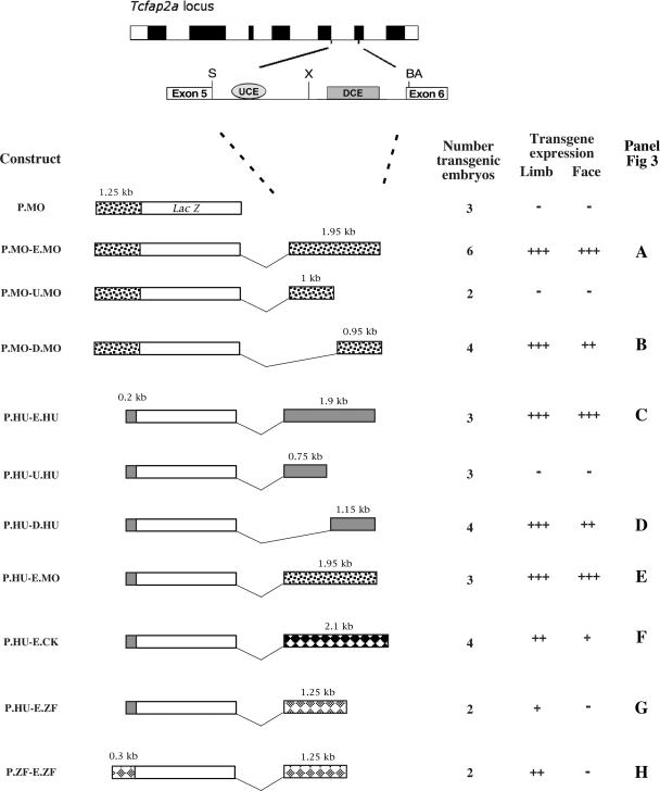
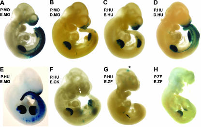
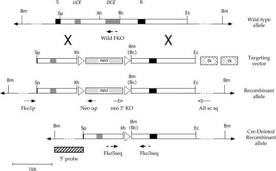
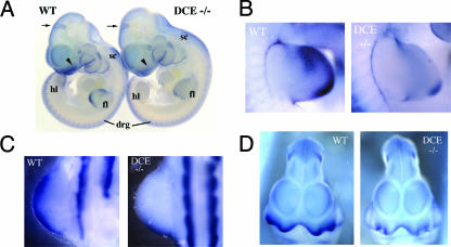
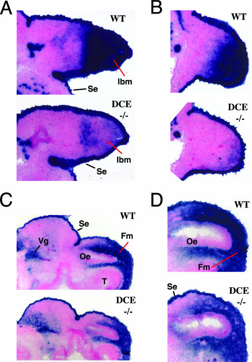
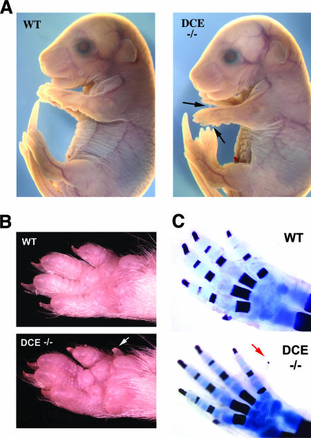
Tcfap2a, the gene encoding the mouse AP-2alpha transcription factor, is required for normal development of multiple structures during embryogenesis, including the face and limbs. Using comparative sequence analysis and transgenic-mouse experiments we have identified an intronic enhancer within this gene that directs expression to the face and limb mesenchyme. There are two conserved sequence blocks within this intron, and the larger of these directs tissue-specific activity and is found in all vertebrate Tcfap2a genes analyzed. To assess the role of the enhancer in regulating endogenous mouse Tcfap2a expression, we have deleted this cis-regulatory sequence from the genome. Loss of this element severely impairs Tcfap2a expression in the limb bud mesenchyme but generates only a modest reduction in the facial mesenchyme. The reduction in Tcfap2a transcription is accompanied by altered patterning of the forelimb, resulting in postaxial polydactyly. These results indicate that the major role for this enhancer resides within the limb bud, and it serves to maintain a level of Tcfap2a expression that limits the size of the hand plate and the associated number of digit primordia. The potential role of this cis-acting sequence in modeling the size and shape of the face and limbs during evolution is discussed.


Figures

FIG. 1.
FIG. 1.
Two conserved sequence blocks occur within the Tcfap2a fifth intron of vertebrates. Shown are sequence alignments of (A) the UCE and (B) the DCE. Identical sequences are boxed, and gaps are indicated by dashes. Numbers above the alignment refer to the positions in the human genomic intron relative to the splice donor of exon 5. Thick gray lines above the sequence indicate the predicted binding sites for the associated transcription factor based on computer prediction programs or experimental analysis (6, 12, 21, 37). Thick black lines indicate the positions of a Sox site and the conserved STAT binding site that is critical for enhancer activity (12). The asterisk in panel B shows the position of a BclI site in the mouse genome that marks the downstream extent of the DCE sequences deleted (see Fig. 4). hu, human; mo, mouse; ck, chicken; xe, frog; zf, zebrafish. bHLH, basic helix-loop-helix; RXR, retinoid X receptor.
FIG. 2.
FIG. 2.
Transgenic analysis of the Tcfap2a FNP/limb bud mesenchyme (LBM) enhancer. (Top) Schematic representation of the Tcfap2a locus with exons depicted as filled boxes. Intron 5 is expanded to highlight two subregions of high sequence conservation, the UCE and the DCE. Abbreviations: S, SpeI; X, XhoI; BA, BsaAI. (Bottom left) Schematic representation of the transgenic constructs utilized in these studies and the associated expression data. Each construct contains basal promoter elements that direct the expression of the Tcfap2a-LacZ fusion transgene. Promoter and enhancer sequences are shown for mice, (dotted rectangles), humans (gray rectangles), chicks (black diamonds), and zebrafish (gray diamonds). The sizes of the fragments are indicated above the rectangles. (Bottom right) Summary of expression data acquired from each transgene. The number of transgenic embryos is indicated for each construct. For constructs such as P.MO that did not yield expression in the face and limbs, this number refers to the number of LacZ-positive embryos obtained in which we saw random and inconsistent patterns of β-galactosidase expression between transgenic embryos. For constructs such as P.MO-E.MO that were expressed in the face and limbs, this number indicates all the transgenic embryos that were obtained and indicates that the locations of β-galactosidase expression were relatively consistent between all embryos. Expression was scored in LBM (limb) and FNP (face). Relative intensity and domain of expression are indicated by the number of plus signs, while a minus sign indicates that no expression was observed in the transgenic embryos. The right column indicates the relevant panel showing a representative expression pattern for the particular construct in Fig. 3.
FIG. 3.
FIG. 3.
Expression of β-galactosidase in transgenic embryos. (A) Lateral views of E10.5 transgenic embryos generated using P.MO-E.MO (A), P.MO-D.MO (B), P.HU-E.HU (C), P.HU-D.HU (D), P.HU-E.MO (E), P.HU-E.CK (F), P.HU-E.ZF (G), and P.ZF-E.ZF (H). Embryos in panels A to E show strong expression in the face and limbs. Panels A, D, and E are examples of embryos with a strong trunk neural crest expression domain. The arrow in panel F shows the limited facial expression domain. The arrow in panel G shows the posterior limb bud expression domain. The asterisk indicates a region of LacZ expression in the central nervous system that was not consistently observed with this transgene. The embryo in panel H was broken prior to photography, but no facial expression was noted in the undamaged intact embryo.
FIG. 4.
FIG. 4.
Derivation of the Tcfap2aDCE−/− allele. Diagrammatic representations of, from top to bottom, wild-type mouse Tcfap2a (top), targeting vector, recombinant locus, and the final DCE-deleted allele after removal of the neo gene with Cre recombinase are shown. Exons 5 and 6 (black boxes), the UCE and DCE (gray boxes), restriction enzyme sites (Bc, BclI; Bm, BamHI; Ec, Eco47III; Sp, SpeI; Xh, XhoI), loxP sites (white triangles) are shown along with neomycin resistance (neo) and thymidine kinase (tk) genes. Note that a novel BamHI site replaces the BclI site, denoted by Bc, located at the 3′ end of the DCE after gene targeting. The positions of the primer pairs used for PCR amplification and genotyping (arrows) and the 5′ probe used for Southern blotting analysis (striped box) are also shown.
FIG. 5.
FIG. 5.
WMISH analysis of Tcfap2a expression in the Tcfap2aDCE−/− mouse. (A) Lateral views of E10.5 wild-type (WT) and Tcfap2aDCE−/− (DCE−/−) embryos stained with a Tcfap2a antisense probe. Arrowheads indicate the FNP, and arrows show domains of expression in the midbrain that are similar in the two embryos. Other regions of Tcfap2a expression that are similar in the two embryos are the trunk NCC-derived dorsal root ganglia (drg) and the spinal cord (sc). Reduced expression is apparent in the forelimb (fl) and hind limb (hl) buds of the Tcfap2aDCE−/− embryo. (B and C) Detailed lateral views of wild-type and Tcfap2aDCE−/− forelimb (B) and hind limb (C) buds. Anterior is to the top. (D) Ventral view of wild-type and Tcfap2aDCE−/− heads.
FIG. 6.
FIG. 6.
In situ analysis of Tcfap2a expression in the Tcfap2aDCE−/− mouse. All sections are stained following hybridization with a Tcfap2a antisense probe. (A and B) Sagittal sections of E10.5 wild-type (WT) and Tcfap2aDCE−/− (DCE−/−) forelimb (A) and hind limb (B) buds. Anterior is to the top. (C) Transverse sections of E10.5 wild-type and Tcfap2aDCE−/− heads. Dorsal is to the left. (D) Detailed view of transverse sections of E10.5 wild-type and Tcfap2aDCE−/− heads. Dorsal is to the left. In panels C and D, note that levels of expression within the surface ectoderms (se) and trigeminal ganglia (Vg) of the two embryos are similar, whereas the intensity of staining is lower in the facial mesenchyme (fm) of the Tcfap2aDCE−/− sample. Abbreviations: lbm, limb bud mesenchyme; oe, olfactory epithelium; t, telencephalon.
FIG. 7.
FIG. 7.
Face and limb morphology in the Tcfap2aDCE−/− mouse. (A) Gross morphology of E16.5 wild-type (WT) and Tcfap2aDCE−/− (DCE−/−) embryos. Both embryos have similar facial appearances, but the Tcfap2aDCE−/− forelimbs have an extra posterior digit nubbin (arrows). (B) Ventral view of forepaws of 6-week-old wild-type and Tcfap2aDCE−/− mice. The extra posterior digit and its associated nail are indicated (arrow). (C) Limb skeletons derived from E18.5 wild-type and Tcfap2aDCE−/− embryos. The extra posterior digit ossification center is shown with an arrow.

References

    1. Ahituv, N., A. Erven, H. Fuchs, K. Guy, R. Ashery-Padan, T. Williams, M. H. de Angelis, K. B. Avraham, and K. P. Steel. 2004. An ENU-induced mutation in AP-2α leads to middle ear and ocular defects in Doarad mice. Mamm. Genome 15424-432. - PubMed
    1. Boffelli, D., M. A. Nobrega, and E. M. Rubin. 2004. Comparative genomics at the vertebrate extremes. Nat. Rev. Genet. 5456-465. - PubMed
    1. Bradshaw, M. S., C. S. Shashikant, H. G. Belting, J. A. Bollekens, and F. H. Ruddle. 1996. A long-range regulatory element of Hoxc8 identified by using the pClasper vector. Proc. Natl. Acad. Sci. USA 932426-2430. - PMC - PubMed
    1. Brewer, S., W. Feng, J. Huang, S. Sullivan, and T. Williams. 2004. Wnt1-Cre mediated deletion of AP-2α causes multiple neural crest related defects. Dev. Biol. 267135-152. - PubMed
    1. Brewer, S., X. Jiang, S. Donaldson, T. Williams, and H. M. Sucov. 2002. Requirement for AP-2α in cardiac outflow tract morphogenesis. Mech. Dev. 110139-149. - PubMed

Publication types

MeSH terms

Substances