. 2007 Nov 26;204(12):2875-88.
doi: 10.1084/jem.20062299. Epub 2007 Nov 12.

Conditional inactivation of Fbxw7 impairs cell-cycle exit during T cell differentiation and results in lymphomatogenesis

Affiliations

Conditional inactivation of Fbxw7 impairs cell-cycle exit during T cell differentiation and results in lymphomatogenesis

Ichiro Onoyama et al. J Exp Med. .

Abstract

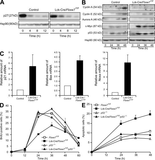
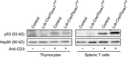
Cell proliferation is strictly controlled during differentiation. In T cell development, the cell cycle is normally arrested at the CD4(+)CD8(+) stage, but the mechanism underlying such differentiation-specific exit from the cell cycle has been unclear. Fbxw7 (also known as Fbw7, Sel-10, hCdc4, or hAgo), an F-box protein subunit of an SCF-type ubiquitin ligase complex, induces the degradation of positive regulators of the cell cycle, such as c-Myc, c-Jun, cyclin E, and Notch. FBXW7 is often mutated in a subset of human cancers. We have now achieved conditional inactivation of Fbxw7 in the T cell lineage of mice and found that the cell cycle is not arrested at the CD4(+)CD8(+) stage in the homozygous mutant animals. The mutant mice manifested thymic hyperplasia as a result of c-Myc accumulation and eventually developed thymic lymphoma. In contrast, mature T cells of the mutant mice failed to proliferate in response to mitogenic stimulation and underwent apoptosis in association with accumulation of c-Myc and p53. These latter abnormalities were corrected by deletion of p53. Our results suggest that Fbxw7 regulates the cell cycle in a differentiation-dependent manner, with its loss resulting in c-Myc accumulation that leads to hyperproliferation in immature T cells but to p53-dependent cell-cycle arrest and apoptosis in mature T cells.


Figures

Figure 1.
Figure 1.
Generation of mice with T cell–specific deficiency of Fbxw7. (A) RT and real-time PCR analysis of the relative abundance of Fbxw7 mRNA in DN, DP, CD4 SP, and CD8 SP mouse thymocytes isolated by flow cytometry. Data are means ± SD of values from three independent experiments. (B) Schematic representations of the wild-type mouse Fbxw7 allele, the targeting vector, the Fbxw7 allele with a loxP-neo cassette (Flox-neo), the floxed Fbxw7 allele (Flox), and the floxed Fbxw7 allele after removal of exon 5 by Cre recombinase (ΔE5). The expected sizes of bands in a Southern blot analysis with probe 1 (for StuI [S] fragments) or probe 2 (for BamHI [B] fragments) are indicated. Exons are shown as numbered closed boxes, and loxP sites are shown as closed triangles. (C) Southern blot analysis with probe 2 of BamHI-digested DNA from the tail of mice of the indicated genotypes. The wild-type and floxed alleles give rise to hybridizing fragments of 7.8 and 1.2 kb, respectively. (D) PCR analysis of genomic DNA from the thymus of Fbxw7F/F, Lck-Cre/Fbxw7+/F, and Lck-Cre/Fbxw7F/F mice with the primers Floxed 1 and Floxed 2 (see Materials and methods). The positions of amplified fragments corresponding to wild-type, floxed, and ΔE5 alleles are indicated.
Figure 2.
Figure 2.
Failure of cell-cycle arrest in DP thymocytes of Lck-Cre/Fbxw7F/Fmice. (A) Gross appearance of the thymus of Lck-Cre/Fbxw7+/F and Lck-Cre/Fbxw7F/F mice at 8 wk of age. Bar, 5 mm. (B) Representative flow cytometric analysis of surface expression of CD4 and CD8 on thymocytes from Lck-Cre/Fbxw7+/F or Lck-Cre/Fbxw7F/F mice at 8 wk of age. The percentages of DN, DP, and SP populations are indicated. (C) Absolute cell numbers for total thymocytes and thymocyte subsets determined as in B. Data are means ± SD of values from 13 Fbxw7F/F (control) and 19 Lck-Cre/Fbxw7F/F mice. **, P < 0.01 using the Student's t test. (D) BrdU incorporation into thymocytes in vivo. The proportion of each thymocyte subset in S phase was determined by measurement of incorporation of BrdU after its intraperitoneal injection in 8-wk-old Lck-Cre/Fbxw7F/F or Fbxw7F/F (control) mice. Data are means ± SD of values from four animals of each genotype. **, P < 0.01 using the Student's t test. (E) Increased expression of Notch1, Notch3, and c-Myc in total thymocytes from Lck-Cre/Fbxw7F/F mice. Total thymocytes of 8-wk-old Lck-Cre/Fbxw7+/F or Lck-Cre/Fbxw7F/F mice were subjected to immunoblot analysis with antibodies to the indicated proteins. (F) Accumulation of Notch1, Notch3, and c-Myc specifically in DP thymocytes of Lck-Cre/Fbxw7F/F mice. Thymocyte subsets of Fbxw7F/F (control) or Lck-Cre/Fbxw7F/F mice were analyzed as in E.
Figure 3.
Figure 3.
c-Myc is responsible for the overproliferation phenotype of Fbxw7-deficient DP thymocytes. (A) Representative flow cytometric analysis of surface expression of CD4 and CD8 on thymocytes from Fbxw7F/F/RBP-JF/F (control), CD4-Cre/Fbxw7F/F, CD4-Cre/Fbxw7F/F/RBP-JF/F, or CD4-Cre/Fbxw7F/F/c-MycF/F mice at 12 wk of age. The respective percentages are indicated. (B) Absolute cell numbers for total thymocytes and thymocyte subsets determined as in A. Data are means ± SD of values from 7 Fbxw7F/F/RBP-JF/F (control), 11 CD4-Cre/Fbxw7F/F, 6 CD4-Cre/Fbxw7F/F/RBP-JF/F, and 5 CD4-Cre/Fbxw7F/F/c-MycF/F mice. *, P < 0.05 using the Student's t test.
Figure 4.
Figure 4.
Lck-Cre/Fbxw7F/Fmice develop T cell lymphoma. (A) Thymi from Fbxw7F/F (control; left) and Lck-Cre/Fbxw7F/F (middle and right) littermates at 14 wk of age. Those from the Lck-Cre/Fbxw7F/F mice were hyperplastic (middle) or tumorous (right). Bar, 5 mm. (B and C) Hematoxylin-eosin staining of sections of the thymus from an Fbxw7F/F mouse (B) or from an Lck-Cre/Fbxw7F/F mouse that developed lymphoma (C). The normal thymic structure was destroyed and replaced with large, atypical lymphoma cells in the latter animal. Bars, 50 μm. (D) Necrotic lesions in the thymus of an Lck-Cre/Fbxw7F/F mouse with lymphoma. Bar, 50 μm. (E and F) Infiltration of lymphoma cells into the fat (E) and lung (F) of Lck-Cre/Fbxw7F/F mice. Bars: (E) 50 μm; (F) 500 μm. (G) Surface expression of CD4 and CD8 on thymic lymphoma cells from an Lck-Cre/Fbxw7F/F mouse. (H) Southern blot analysis of genomic DNA from the tail and thymus of wild-type mice and from two thymic lymphomas of Lck-Cre/Fbxw7F/F mice. The DNA was digested with BamHI and probed with a 2.1-kb EcoRI fragment of the TCR-Jβ2 locus. The positions of germline and rearranged (arrowheads) fragments are indicated. (I) Kaplan-Meier plot of the overall survival of mice of the indicated genotypes.
Figure 5.
Figure 5.
Proliferative defect of Fbxw7-deficient mature T cells. (A) Representative flow cytometric analysis of surface expression of TCRβ and B220 (top) or of CD4 and CD8 (bottom) on spleen cells from Fbxw7F/F (control) or Lck-Cre/Fbxw7F/F mice at 8 wk of age. The respective percentages are indicated. (B) Absolute cell numbers of splenocyte subsets determined as in A. Data are means ± SD of values from 8 Fbxw7F/F (control) and 14 Lck-Cre/Fbxw7F/F mice. ***, P < 0.005 using the Student's t test. (C) Splenic T cells from Lck-Cre/Fbxw7+/F (control) or Lck-Cre/Fbxw7F/F mice were stimulated for 48 h with the indicated concentrations of anti-CD3ε (left) or ionomycin and phorbol 12,13-dibutyrate (PDBu; right), after which the incorporation of [3H]thymidine was measured. Data are means ± SD of values from three independent experiments. (D) Splenic T cells from Fbxw7F/F (control) or Lck-Cre/Fbxw7F/F mice were left unstimulated (open trace) or stimulated with 5 μg/ml anti-CD3ε for 7 h (closed trace) and were then subjected to flow cytometric analysis for detection of the early activation antigen CD69. (E) Immunoblot analysis of phospho-Lck and Lck (loading control), phospho-ERK and ERK (loading control), and phospho-JNK and JNK (loading control) in splenic T cells from Fbxw7F/F (control) or Lck-Cre/Fbxw7F/F mice at 0, 5, and 10 min after stimulation with 5 μg/ml anti-CD3ε. The intensity of the bands corresponding to the phosphorylated proteins was normalized by that of total proteins. The value at time = 0 is defined as 1.
Figure 6.
Figure 6.
Abnormal accumulation of c-Myc and induction of p53 in stimulated splenic T cells from Lck-Cre/Fbxw7F/F mice. (A) Immunoblot analysis of p27 and Hsp90 (loading control) in splenic T cells from Fbxw7F/F (control) or Lck-Cre/Fbxw7F/F mice at 0, 4, 8, and 12 h after stimulation with 5 μg/ml anti-CD3ε. (B) Immunoblot analysis of cyclin A, cyclin E, Aurora A, c-Myc, p53, and Hsp90 in splenic T cells from Fbxw7F/F (control) or Lck-Cre/Fbxw7F/F mice at 0, 24, 36, and 48 h after stimulation with 5 μg/ml anti-CD3ε. (C) RT and real-time PCR analysis of p53-dependent gene expression in splenic T cells from Fbxw7F/F (control) or Lck-Cre/Fbxw7F/F mice after stimulation for 24 h with 5 μg/ml anti-CD3ε. Normalized data for p21, Bax, and Noxa mRNAs are expressed relative to the corresponding values for cells from control mice and are means ± SD of values from three independent experiments. (D) Splenic T cells from mice of the indicated genotypes were stimulated with 5 μg/ml anti-CD3ε for the indicated times and exposed to BrdU during the final 1 h of incubation. They were then stained with anti-BrdU, and the percentage of BrdU-positive cells was determined by flow cytometry. Data are means ± SD of values from three independent experiments. (E) Splenic T cells stimulated as in D were stained with propidium iodide, and the percentage of sub-G1 (apoptotic) cells was determined by flow cytometry. Data are means ± SD of values from three independent experiments.
Figure 7.
Figure 7.
Exaggerated induction of p53 in splenic T cells from Lck-Cre/Fbxw7F/F mice. Thymocytes and splenic T cells isolated from Fbxw7F/F (control) or Lck-Cre/Fbxw7F/F mice were incubated in the absence or presence of 5 μg/ml anti-CD3ε for 24 h, after which cell lysates were subjected to immunoblot analysis with antibodies to p53 and to Hsp90 (loading control).

References

    1. Conlon, I., and M. Raff. 1999. Size control in animal development. Cell. 96:235–244. - PubMed
    1. Eilers, M., S. Schirm, and J.M. Bishop. 1991. The MYC protein activates transcription of the alpha-prothymosin gene. EMBO J. 10:133–141. - PMC - PubMed
    1. Blackwood, E.M., L. Kretzner, and R.N. Eisenman. 1992. Myc and Max function as a nucleoprotein complex. Curr. Opin. Genet. Dev. 2:227–235. - PubMed
    1. Yada, M., S. Hatakeyama, T. Kamura, M. Nishiyama, R. Tsunematsu, H. Imaki, N. Ishida, F. Okumura, K. Nakayama, and K.I. Nakayama. 2004. Phosphorylation-dependent degradation of c-Myc is mediated by the F-box protein Fbw7. EMBO J. 23:2116–2125. - PMC - PubMed
    1. Welcker, M., A. Orian, J. Jin, J.A. Grim, J.W. Harper, R.N. Eisenman, and B.E. Clurman. 2004. The Fbw7 tumor suppressor regulates glycogen synthase kinase 3 phosphorylation-dependent c-Myc protein degradation. Proc. Natl. Acad. Sci. USA. 101:9085–9090. - PMC - PubMed

MeSH terms