. 2007 Nov 26;204(12):2889-97.
doi: 10.1084/jem.20071283. Epub 2007 Nov 5.

An ITAM-signaling pathway controls cross-presentation of particulate but not soluble antigens in dendritic cells

Affiliations

An ITAM-signaling pathway controls cross-presentation of particulate but not soluble antigens in dendritic cells

Daniel B Graham et al. J Exp Med. .

Abstract

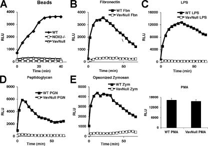
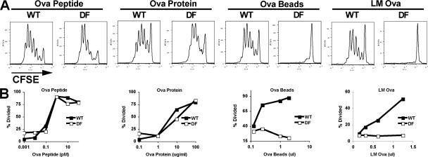
Dendritic cells (DC) possess a unique capacity for presenting exogenous antigen on major histocompatibility class I, a process that is referred to as cross-presentation, which serves a critical role in microbial and tumor immunity. During cross-presentation, antigens derived from pathogen-infected or tumor cells are internalized and processed by DCs for presentation to cytotoxic T lymphocytes (CTLs). We demonstrate that a signaling pathway initiated by the immunoreceptor tyrosine-based activation motif (ITAM)-containing adaptors DAP12 and FcRgamma utilizes the Vav family of Rho guanine nucleotide exchange factors (GEFs) for processing and cross-presentation of particulate, but not soluble, antigens by DCs. Notably, this novel pathway is crucial for processing and presentation of particulate antigens, such as those associated with Listeria monocytogenes bacteria, yet it is not required for antigen uptake. Mechanistically, we provide evidence that in DCs, Vav GEFs are essential to link ITAM-dependent receptors with the activation of the NOX2 complex and production of reactive oxygen species (ROS), which regulate phagosomal pH and processing of particulate antigens for cross-presentation. Importantly, we show that genetic disruption of the DAP12/FcRgamma-Vav pathway leads to antigen presentation defects that are more profound than in DCs lacking NOX2, suggesting that ITAM signaling also controls cross-presentation in a ROS-independent manner.


Figures

Figure 1.
Figure 1.
Vav is required for cross-presentation of particulate, but not soluble, antigen. (A) WT and VavNULL BMDCs were cultured with CFSE-labeled OT-1 T cells and the indicated antigens for 72 h. T cell proliferation was determined by CFSE dye dilution and FACS analysis. (B) T cell proliferation was determined as in A and plotted as antigen dose versus the percentage of T cells that had undergone at least one division.
Figure 2.
Figure 2.
Vav is not required for macropinocytosis of soluble antigen. (A) BMDCs were cultured with 70 kD FITC-Dextran (FITC-Dex) for the indicated time points at 4 or 37°C. FITC uptake by macropinocytosis was determined by FACS. (B) BMDCs were treated as in A and examined for FITC uptake by single-cell imaging. Bar, 5 μm.
Figure 3.
Figure 3.
Vav is not required for phagocytosis of latex beads or bacteria. (A) BMDCs were stained with FITC anti-CD11b to demarcate the membrane and cultured with latex beads. Cells were then distributed onto slides and analyzed by confocal microscopy. Optical slices through the z plane were imaged at 2-μm increments from the bottom of the cell to the top, and they are portrayed from left to right. DIC images clearly reveal the bead, which is highly refractive. Bar, 5 μm. (B) BMDCs were cultured with Alexa Fluor 647-labeled latex beads and analyzed for phagocytosis by FACS. (C) BMDCs were cultured with latex beads for the indicated time points and scored for phagocytosis by microscopy. Data represent the mean ± the SD of the percentage of cells that internalized at least one bead. Scoring was performed in triplicate and accounted for at least 125 cells per condition. (D) BMDCs were cultured with CFSE-labeled LM for the indicated time points at 37°C. The percentage of cells that had internalized at least one bacterium is expressed as the mean ± the SD of triplicate scoring, which included at least 125 cells per condition.
Figure 4.
Figure 4.
Vav is required for ROS production in DCs. ROS production in BMDCs was determined with the chemiluminescent substrate lucigenin. Cells were stimulated with 5 μl/sample latex beads (A), 1 μg/ml plate-bound fibronectin (Fbn; B), 10 μg/ml LPS (C), 10 μg/ml peptidoglycan (PGN; D), and 20 μg/ml zymosan (Zym; E). (inset) Cells stimulated with 50 ng/ml PMA for 60-70 min. RLU, relative light units.
Figure 5.
Figure 5.
Vav controls phagosomal pH and antigen degradation. (A) Phagosomal pH was measured in BMDCs using latex beads coupled with pH-sensitive FITC and pH-insensitive Alexa Fluor 647. DCs were loaded with beads for 30 min, washed, and cultured for the indicated time points before analysis by FACS. For analysis, cells that had internalized equal numbers of beads (based on Alexa Fluor 647 MFI) were gated and analyzed for FITC MFI. Data represent the relative fold change in FITC MFI over time. (B) Antigen degradation in phagosomes was monitored using latex beads covalently coupled to OVA. DCs were loaded with beads for 30 min, washed, and cultured for the indicated time points. Beads were then recovered by lysing the cells and stained with anti-OVA antibodies before FACS analysis.
Figure 6.
Figure 6.
Adhesion-dependent Vav phosphorylation and ROS production in DCs requires DAP12 and FcRγ. (A) WT and DAP12 and FcRγ (DF) BMDCs were stimulated on fibrinogen-coated plates (150 μg/ml) and lysed at the indicated time points. Vav was immunoprecipitated from the lysates, resolved by SDS-PAGE, and detected by Western blot with anti-phospho-tyrosine antibody. Blots were subsequently stripped and reprobed with anti-Vav antibody to demonstrate equal protein loading. (B) Alternatively, whole-cell lysates were blotted for phospho-ERK, stripped, and reprobed for total ERK. (C) ROS production in WT and DF BMDCs was determined with the chemiluminescent substrate lucigenin. Cells were stimulated with latex beads, LM, or LPS (10 μg/ml). RLU, relative light units.
Figure 7.
Figure 7.
DAP12 and FcRγ are required for cross-presentation of particulate antigens. (A) CFSE-labeled OT-1 T cells were cultured with WT and DAP12−/−FcRγ−/− (DF) BMDCs, along with the indicated antigens. Cells were stimulated for 3 d before analysis of T cell proliferation by CFSE dye dilution and FACS. (B) T cell proliferation was determined as in Fig. 6 B and plotted as antigen dose versus the percentage of T cells that had undergone at least one division.

References

    1. Bevan, M.J. 1976. Cross-priming for a secondary cytotoxic response to minor H antigens with H-2 congenic cells which do not cross-react in the cytotoxic assay. J. Exp. Med. 143:1283–1288. - PMC - PubMed
    1. Lanzavecchia, A. 1996. Mechanisms of antigen uptake for presentation. Curr. Opin. Immunol. 8:348–354. - PubMed
    1. Hall, A.B., M.A. Gakidis, M. Glogauer, J.L. Wilsbacher, S. Gao, W. Swat, and J.S. Brugge. 2006. Requirements for Vav guanine nucleotide exchange factors and Rho GTPases in FcgammaR- and complement-mediated phagocytosis. Immunity. 24:305–316. - PubMed
    1. Rock, K.L., and L. Shen. 2005. Cross-presentation: underlying mechanisms and role in immune surveillance. Immunol. Rev. 207:166–183. - PubMed
    1. Bevan, M.J. 2006. Cross-priming. Nat. Immunol. 7:363–365. - PubMed

Publication types

MeSH terms

Substances