. 2008 May;115(5):509-32.
doi: 10.1007/s00401-007-0311-9. Epub 2007 Nov 6.

Twenty-first century brain banking. Processing brains for research: the Columbia University methods

Affiliations

Twenty-first century brain banking. Processing brains for research: the Columbia University methods

Jean Paul G Vonsattel et al. Acta Neuropathol. 2008 May.

Abstract

Carefully categorized postmortem human brains are crucial for research. The lack of generally accepted methods for processing human postmortem brains for research persists. Thus, brain banking is essential; however, it cannot be achieved at the cost of the teaching mission of the academic institution by routing brains away from residency programs, particularly when the autopsy rate is steadily decreasing. A consensus must be reached whereby a brain can be utilizable for diagnosis, research, and teaching. The best diagnostic categorization possible must be secured and the yield of samples for basic investigation maximized. This report focuses on integrated, novel methods currently applied at the New York Brain Bank, Columbia University, New York, which are designed to reach accurate neuropathological diagnosis, optimize the yield of samples, and process fresh-frozen samples suitable for a wide range of modern investigations. The brains donated for research are processed as soon as possible after death. The prosector must have a good command of the neuroanatomy, neuropathology, and the protocol. One half of each brain is immersed in formalin for performing the thorough neuropathologic evaluation, which is combined with the teaching task. The contralateral half is extensively dissected at the fresh state. The anatomical origin of each sample is recorded using the map of Brodmann for the cortical samples. The samples are frozen at -160 degrees C, barcode labeled, and ready for immediate disbursement once categorized diagnostically. A rigorous organization of freezer space, coupled to an electronic tracking system with its attached software, fosters efficient access for retrieval within minutes of any specific frozen samples in storage. This report describes how this achievement is feasible with emphasis on the actual processing of brains donated for research.


Figures

Fig. 1
Fig. 1
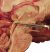
Usual variant for sectioning the brain sagittally. The dorsoventral sagittal cut through the corpus callosum is the simplest variant; however, it requires experience to obtain a cut passing exactly through the midline (a). To secure a section passing perfectly through the midline, four sheets of gauze are rolled together along their longitudinal axis, stretched, and carefully apposed to the dorsal aspect of the corpus callosum (b). The brain is rotated so that its dorsal aspect is resting on the cutting board. Then the knife is carefully placed along the midline of the brainstem and between the frontal lobes, ventrally, and then the cut proceeds, possibly with one stroke, using the full length of the blade. Note the presence of a lipoma attached to the right, lateral aspect of the infundibulum (c). Medial aspect of the left half-brain sectioned in a ventrodorsal direction after having placed the gauze belt over the corpus callosum (d)
Fig. 2
Fig. 2
Variant applied when processing brains of patients with amyotrophic lateral sclerosis, which is referred to as “ALS variant of Protocol 1” (P1-ALS variant). This variant secures the presence of the bilateral nucleus of the hypoglossal nerve (XII) in blocks used either for research (frozen) or diagnostic (formalin fixed). The myelencephalon (a) is detached from the metencephalon (b) before the sagittal cut through the corpus callosum (c). Then 0.3 cm-thick transverse slices are obtained from the myelencephalon. Alternate slices are frozen (d) or fixed (not shown)
Fig. 3
Fig. 3
One half-brain is processed fresh. Up to 150 blocks and parenchymal aliquots are harvested and barcode labeled (de-identified link and site of origin). The contralateral half is fixed for neuropathological evaluations
Fig. 4
Fig. 4
Standard brain blocks (SBB): a series of 36 blocks are obtained from 18 precisely selected areas of each half-brain (see Figs. 12, 22). Representative, frozen blocks (SBB4, 5, 6), obtained from the fresh half-brain (upper right) or from the fixed half-brain (middle right), from which Luxol fast blue counterstained with hematoxylin and eosin 7 μm thick sections are produced for microscopic evaluation (lower right), are perfectly matched. See text
Fig. 5
Fig. 5
Alternate, fresh, and LNV-frozen hemi mesencephalon: left, rostral fresh/frozen slices; right, caudal transverse fresh/frozen slices, each about 0.3 cm thick. The frozen slices are resting on a cold Teflon-coated aluminum plate (see text). The rostral level is especially suitable for research focusing on progressive supranuclear palsy (PSP), and the caudal level for studies on Parkinson’s disease (PD)
Fig. 6
Fig. 6
Separation of the hemi brainstem with attached cerebellum from the cerebral hemisphere. The plan of the cut grazes the ventral edge of the mammillary body, and the dorsal edge of the superior colliculus
Fig. 7
Fig. 7
Slide holder with gauze and hemi brainstem, the cut surface of which is applied against the bottom or lid of the stabilized slide holder to obtain regular, transverse, 0.3 cm thick slices. The razor blade and the vertical aluminum plate keep the slide holder steady
Fig. 8
Fig. 8
The posterior aspect of the cutting ice support (CIS) includes two hooks (framed) to which the carpet of gauze is anchored; the vertical plate on the back stabilizes the CIS (a). Anterior aspect of the CIS (b): first, an aluminum plate is apposed vertically against the front of the ice-filled container to provide a perfect flat surface. Then, one end of the carpet of gauze is anchored to one hook, which is on the posterior aspect of the CIS (a). The carpet is stretched over the aluminum plate on the front, and anchored to the other hook of the CIS on the back. Thus, the stretched carpet evenly covers the aluminum plate apposed against the anterior aspect of the ice-filled container. A rubber band braces the carpet to steady it. Lateral view of the CIS (c). The CIS is resting on a cork cutting board and is apposed against the vertical aluminum plate, which is on the back (a). The cerebral hemisphere is placed on the movable plastic cutting board that abuts the front of the CIS and is slightly elevated to take full advantage of the vertical, carpeted surface (c). The long axis of the movable cutting board and the tangent of the ventral aspect of both the temporal and occipital lobes are aligned and strictly perpendicular to the anterior aspect of the CIS. These axes, the vertical carpeted surface of the CIS, and the long axis of the cork cutting board serve as guides while sectioning the cerebral hemisphere. The tangent to the ventral aspect of the temporal and occipital lobes of a right cerebral hemisphere is aligned with the long edge of the movable cutting board, and is near the right-handed prosector; the blade of the knife is parallel to the carpeted surface (d); conversely, the tangent is away from the right-handed prosector when a left cerebral hemisphere is processed (e). The long edges of the movable cutting board are kept perpendicular to the CIS and the cut surface of the hemisphere is evenly applied against the carpet (e)
Fig. 9
Fig. 9
The Brodmann areas (BA) are shown color-coded on the lateral aspect of the left cerebral hemisphere
Fig. 10
Fig. 10
Posterior aspects of fresh, coronal slices of two distinct hemispheres passing through the amygdala, as they appear on the cold plate (Fig. 11), ready for the harvest of either the standardized (SBB) or additional blocks (ABB), and of the aliquots (vials). The Brodmann areas (BA) are shown color-coded (a) and are used as one of the identifiers of the blocks that include the cortex. The framed areas represent the standardized brain blocks SBB8 (amygdala), SBB7 (GP, globus pallidus), and SBB16 (cingulate gyrus) (b)
Fig. 11
Fig. 11
The cold surface consists of a Styrofoam platform on a mobile cart, which is overlaid by a carpet of ice packs wrapped with a cotton towel on top of which a steel plate is placed. The space between the ice packs and the steel plate can be adjusted to control the temperature of the plate. On the cold steel plate, the coronal slices of the cerebral hemisphere are laid down with the posterior aspect faced up. The container on the left corner of the steel plate holds absolute ethyl alcohol and a razor blade for harvesting blocks from the coronal slices
Fig. 12
Fig. 12
Posterior aspect of fresh, cerebral hemispheric slices, rostral aspect of transverse mesencephalic and myelencephalic slices, and sagittal cerebellar slice. The telencephalic, diencephalic, and cerebellar areas are framed from which the standard brain blocks (SBB) are obtained. The mesencephalon yields two blocks in toto (SBB10.1, 10.2), the myelencephalon two to four (SBB13.1, etc.), as does the metencephalon (not shown)
Fig. 13
Fig. 13
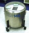
Vessel model XLC140, and a pair of Teflon-coated aluminum plates. A pair of Teflon-coated, aluminum plates (each plate measuring 1.0 × 8.0 × 10.0 cm) for freezing blocks of tissue, rest on the shoulder of the double-walled vacuum vessel XLC140 (XLC140 vessel provider (8/2002): LNV Freezing Vessel, MVE, Inc., Two Appletree Square, Suite 100, 8011 34th Avenue South, Bloomington, MN 55425-1636. Phone: 1-800-2474446, ask for Steve Shaw, Sales Manager. XLC140 vessel with plain lid cover, foam insulation, vapor shipper insert, and caster wheelbase. Ask for Harvard BTRC specifications.)
Fig. 14
Fig. 14
Lower Teflon-coated aluminum plate with fresh frozen samples, as they appear upon removal of the upper Teflon-coated aluminum plate. Five levels of the frozen hippocampal formation (SBB5, body; SBB18, head of hippocampus) were obtained from one half-brain (a). Lower plate with 24 alternate frozen segments of the spinal cord (b)
Fig. 15
Fig. 15
Operational and storage freezers; sample dispersal, CO2 backups, and alarm. Each freezer is connected to: (1) the standard hospital electric supply network; (2) the emergency generator dependent electrical supply network; (3) a CO2 cylinder; and (4) remote alarm system. An extra backup set of CO2 cylinders is kept in the storage freezer room
Fig. 16
Fig. 16
Empty SBB, or ABB, or block “box-area” (Fiberboard Storage Box, Fisher catalogue 5954, divider 11-678-24C) (a). Partially filled SBB, or ABB, or block “box-area” (b)
Fig. 17
Fig. 17
Empty vial “box-area” (Fiberboard Storage Box, Fisher catalogue 5954, divider 13-989-218) (a). Filled vial “box-area.” The filled vial “box-area” contains 81 vials, each of which is electronically tracked (b)
Fig. 18
Fig. 18
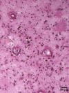
Cerebral cortex [SBB4, Brodmann area 18; 60-year-old man, control; postmortem interval frozen (PMI-fzn): 14:30 hours] frozen with liquid nitrogen vapor. The cortico-subcortical junction is at the bottom of the picture (a). The field magnified (b) is from an area located near the center of a, just to the left of the V-shaped vessel. The morphology of neurons, astrocytes, or oligodendrocytes, is preserved. A 10 μm thick section. HE
Fig. 19
Fig. 19
Cerebral cortex [SBB3, Brodmann area 7; 91-year-old demented woman with multiple cerebral hemorrhages; postmortem interval frozen (PMI-fzn): 17:45 hours] frozen with liquid nitrogen vapor. Freezing artifacts are minimal despite the poor condition of the brain in the fresh state. Cerebral amyloid angiopathy, hemosiderinophages, gliosis, neuritic plaques, and neuronal loss are clearly identifiable. A 10 μm thick section. HE
Fig. 20
Fig. 20
Cerebral cortex [SBB4, Brodmann area 18; 92-year-old demented woman; postmortem interval frozen (PMI-fzn): 03:55 hours] frozen with dry ice. Marked ice-artifacts; however, cellular identification is still possible. A 10 μm thick section. LHE
Fig. 21
Fig. 21
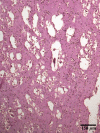
Serial, 10 μm thick sections from the cerebral cortex (same patient as in Fig. 19) without (a) or with (b) prominent artifacts, which consist of innumerable, optically empty vacuoles. These vacuoles are not due to the original freezing of the block, as they are not seen in a or in Fig. 19, but secondary to dehydration that occurred during the interval of time that the thin section was laid on the slide and immersed in absolute alcohol. HE
Fig. 22
Fig. 22
Eighteen standardized blocks harvested for microscopic examination. This series matches the standardized series obtained from the contralateral half-brain prepared for research, which are referred to as standard brain blocks (SBB)

Comment in

Similar articles

Cited by

References

    1. {'text': '', 'ref_index': 1, 'ids': [{'type': 'DOI', 'value': '10.1111/j.1471-4159.1979.tb04607.x', 'is_inner': False, 'url': 'https://doi.org/10.1111/j.1471-4159.1979.tb04607.x'}, {'type': 'PubMed', 'value': '34670', 'is_inner': True, 'url': 'https://pubmed.ncbi.nlm.nih.gov/34670/'}]}
    2. Albrecht J, Yanagihara T (1979) Effect of anoxia and ischemia on ribonuclease activity in brain. J Neurochem 32:1131–1133 - PubMed
    1. {'text': '', 'ref_index': 1, 'ids': [{'type': 'PMC', 'value': 'PMC6578676', 'is_inner': False, 'url': 'https://pmc.ncbi.nlm.nih.gov/articles/PMC6578676/'}, {'type': 'PubMed', 'value': '8774439', 'is_inner': True, 'url': 'https://pubmed.ncbi.nlm.nih.gov/8774439/'}]}
    2. Anderson AJ, Su JH, Cotman CW (1996) DNA damage and apoptosis in Alzheimer’s disease: colocalization with c-Jun immunoreactivity, relationship to brain area, and effect of postmortem delay. J Neurosci 16:1710–1719 - PMC - PubMed
    1. {'text': '', 'ref_index': 1, 'ids': [{'type': 'PMC', 'value': 'PMC1083246', 'is_inner': False, 'url': 'https://pmc.ncbi.nlm.nih.gov/articles/PMC1083246/'}, {'type': 'PubMed', 'value': '125784', 'is_inner': True, 'url': 'https://pubmed.ncbi.nlm.nih.gov/125784/'}]}
    2. Aquilonius SM, Eckernäs SA, Sundwall A (1975) Regional distribution of choline actyltransferase in the human brain: changes in Huntington’s chorea. J Neurol Neurosurg Psychiatry 38:669–677 - PMC - PubMed
    1. {'text': '', 'ref_index': 1, 'ids': [{'type': 'DOI', 'value': '10.1016/S0891-0618(01)00099-0', 'is_inner': False, 'url': 'https://doi.org/10.1016/s0891-0618(01)00099-0'}, {'type': 'PubMed', 'value': '11470556', 'is_inner': True, 'url': 'https://pubmed.ncbi.nlm.nih.gov/11470556/'}]}
    2. Bahn S, Augood SJ, Ryan M, Standaert DG, Starkey M, Emson PC (2001) Gene expression profiling in the post-mortem human brain––no cause for dismay. J Chem Neuroanat 22:79–94 - PubMed
    1. {'text': '', 'ref_index': 1, 'ids': [{'type': 'DOI', 'value': '10.1111/j.1471-4159.1993.tb03532.x', 'is_inner': False, 'url': 'https://doi.org/10.1111/j.1471-4159.1993.tb03532.x'}, {'type': 'PubMed', 'value': '7685811', 'is_inner': True, 'url': 'https://pubmed.ncbi.nlm.nih.gov/7685811/'}]}
    2. Barton AJ, Pearson RC, Najlerahim A, Harrison PJ (1993) Pre- and postmortem influences on brain RNA. J Neurochem 61:1–11 - PubMed

Publication types