

Functional characteristics of patients with retinal dystrophy that manifest abnormal parafoveal annuli of high density fundus autofluorescence; a review and update
- PMID: 17985165
- PMCID: PMC2244701
- DOI: 10.1007/s10633-007-9087-4
Functional characteristics of patients with retinal dystrophy that manifest abnormal parafoveal annuli of high density fundus autofluorescence; a review and update
Abstract
Purpose: To examine the presence and functional significance of annular fundus autofluorescence abnormalities in patients with different retinal dystrophies.
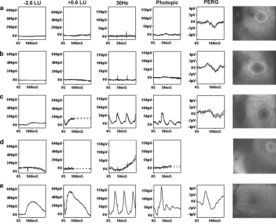
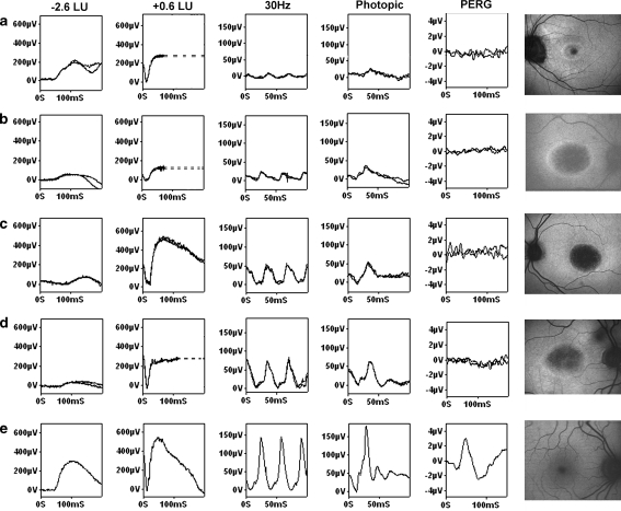
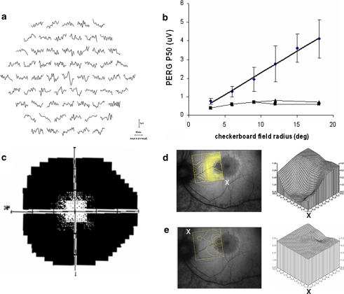
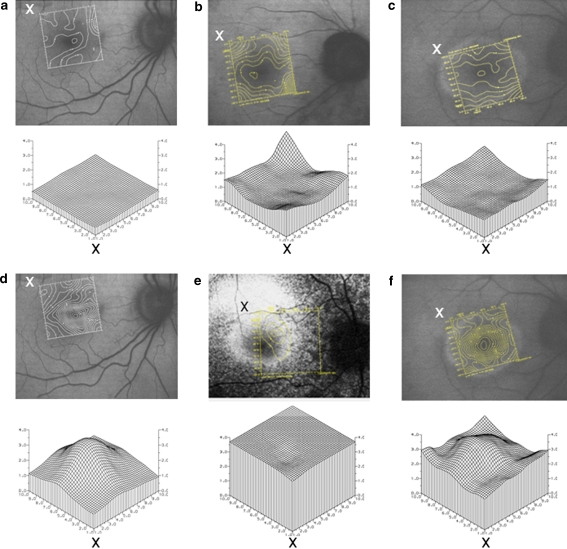
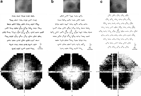
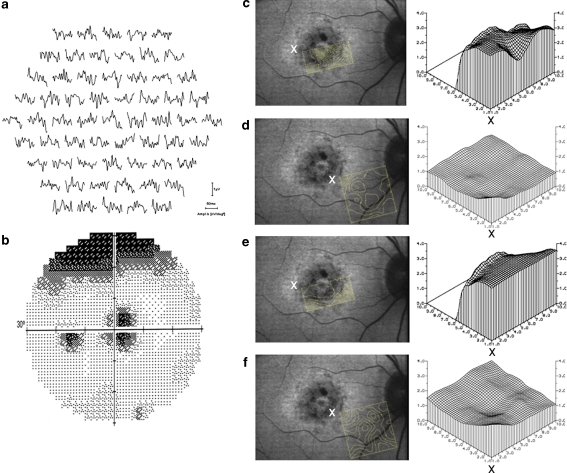
Methods: Eighty one patients were ascertained who had a parafoveal ring of high density on fundus autofluorescence imaging. Sixty two had had a clinical diagnosis of retinitis pigmentosa (RP) or Usher syndrome with normal visual acuity. Others included a case of Leber congenital amaurosis and genetically confirmed cases of cone or cone-rod dystrophy (GUCA1A, RPGR, RIMS1), "cone dystrophy with supernormal rod ERG" (KCNV2) and X-linked retinoschisis (RS1). International-standard full-field and pattern electroretinography (ERG; PERG) were performed. Some patients with rod-cone or cone-rod dystrophy underwent multifocal ERG (mfERG) testing and photopic and scotopic fine matrix mapping (FMM).
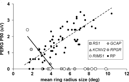
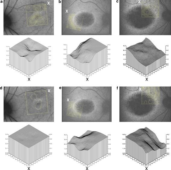
Results: In patients with RP, the radius of the parafoveal ring of high density correlated with PERG P50 (R = 0.83, P < 0.0005, N = 62) and encircled areas of preserved photopic function. In the other patients, AF rings either resembled those seen in RP or encircled an area of central atrophy. Ring radius was inversely related to the PERG P50 component in 4 of 18 cases with a detectable response. FMM showed that arcs of high density were associated with a gradient of sensitivity change.
Conclusions: Parafoveal rings of high density autofluorescence are a non-specific manifestation of retinal dysfunction that can occur in different retinal dystrophies. Electrophysiology remains essential for accurate diagnosis. The high correlation of autofluorescence with PERG, mfERG and FMM demonstrates that AF abnormalities have functional significance and may help identify suitable patients and retinal areas amenable to future therapeutic intervention.
Figures
References
-
- {'text': '', 'ref_index': 1, 'ids': [{'type': 'PubMed', 'value': '669891', 'is_inner': True, 'url': 'https://pubmed.ncbi.nlm.nih.gov/669891/'}]}
- Wing GL, Blanchard GC, Weiter JJ (1978) The topography and age relationship of lipofuscin concentration in the retinal pigment epithelium. Invest Ophthalmol Vis Sci 17:601–607 - PubMed
-
- {'text': '', 'ref_index': 1, 'ids': [{'type': 'PubMed', 'value': '6589859', 'is_inner': True, 'url': 'https://pubmed.ncbi.nlm.nih.gov/6589859/'}]}
- Feeney-Burns L, Eldred GE (1983) The fate of the phagosome: conversion to “Age Pigment” and impact in human retinal pigment epithelium. Trans Ophthal Soc UK 103:416–421 - PubMed
-
- {'text': '', 'ref_index': 1, 'ids': [{'type': 'PubMed', 'value': '8849547', 'is_inner': True, 'url': 'https://pubmed.ncbi.nlm.nih.gov/8849547/'}]}
- Kennedy CJ, Rakoczy PE (1995) Constable IJ. Lipofuscin of the retinal pigment epithelium: a review. Eye 9:763–771 - PubMed
-
- {'text': '', 'ref_index': 1, 'ids': [{'type': 'PubMed', 'value': '11431454', 'is_inner': True, 'url': 'https://pubmed.ncbi.nlm.nih.gov/11431454/'}]}
- Delori FC, Goger DG, Dorey CK (2001) Age-related accumulation and spatial distribution of lipofuscin in RPE of normal subjects. Invest Ophthalmol Vis Sci. 42:1855–1866 - PubMed
-
- {'text': '', 'ref_index': 1, 'ids': [{'type': 'DOI', 'value': '10.1136/bjo.79.5.407', 'is_inner': False, 'url': 'https://doi.org/10.1136/bjo.79.5.407'}, {'type': 'PMC', 'value': 'PMC505125', 'is_inner': False, 'url': 'https://pmc.ncbi.nlm.nih.gov/articles/PMC505125/'}, {'type': 'PubMed', 'value': '7612549', 'is_inner': True, 'url': 'https://pubmed.ncbi.nlm.nih.gov/7612549/'}]}
- von Rückmann A, Fitzke FW, Bird AC (1995) Distribution of fundus autofluorescence with a scanning laser ophthalmoscope. Br J Ophthalmol 79:407–412 - PMC - PubMed
Publication types
MeSH terms
Substances
LinkOut - more resources
Full Text Sources
Medical
Molecular Biology Databases
Research Materials
