. 2008 Mar 1;122(5):978-89.
doi: 10.1002/ijc.23221.

Prevention of upper aerodigestive tract cancer in zinc-deficient rodents: inefficacy of genetic or pharmacological disruption of COX-2

Affiliations

Prevention of upper aerodigestive tract cancer in zinc-deficient rodents: inefficacy of genetic or pharmacological disruption of COX-2

Louise Y Y Fong et al. Int J Cancer. .

Abstract

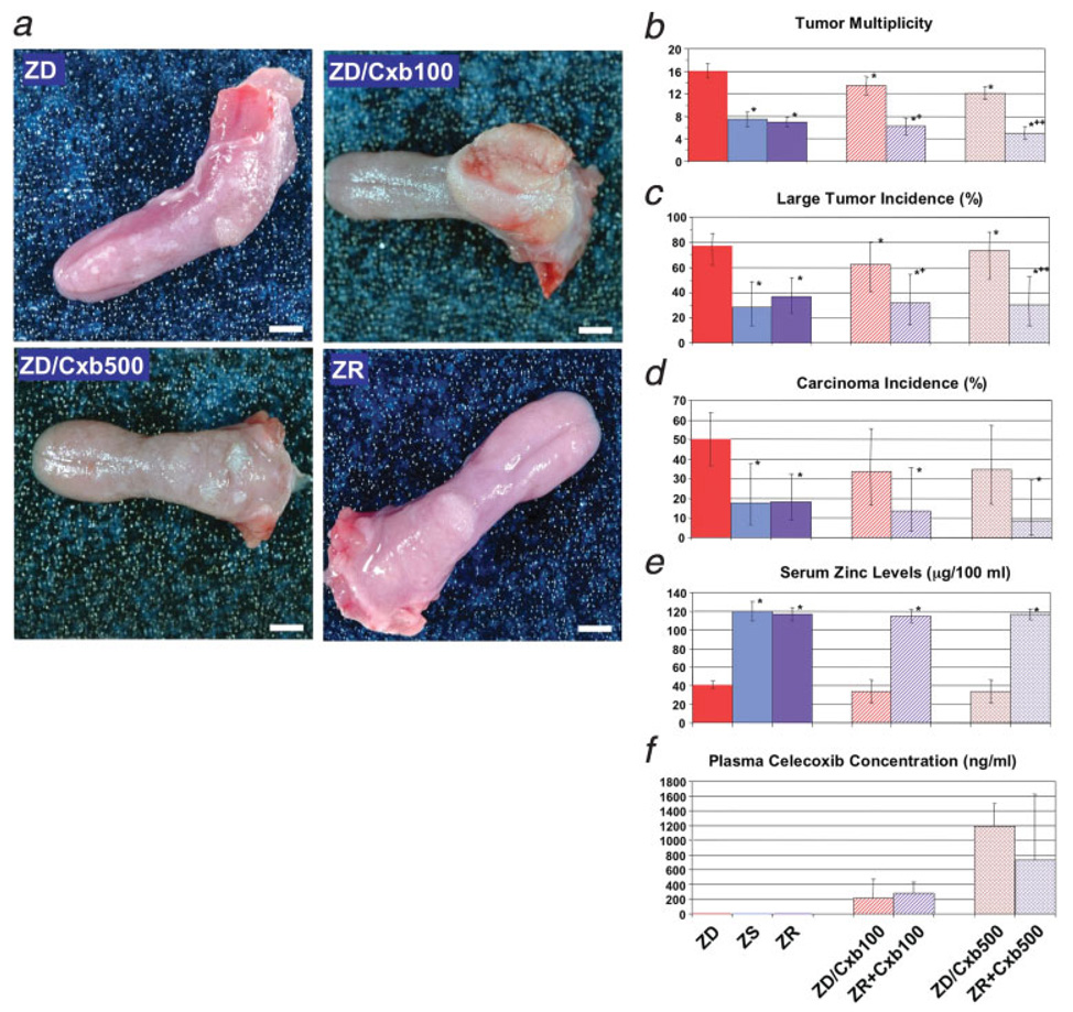
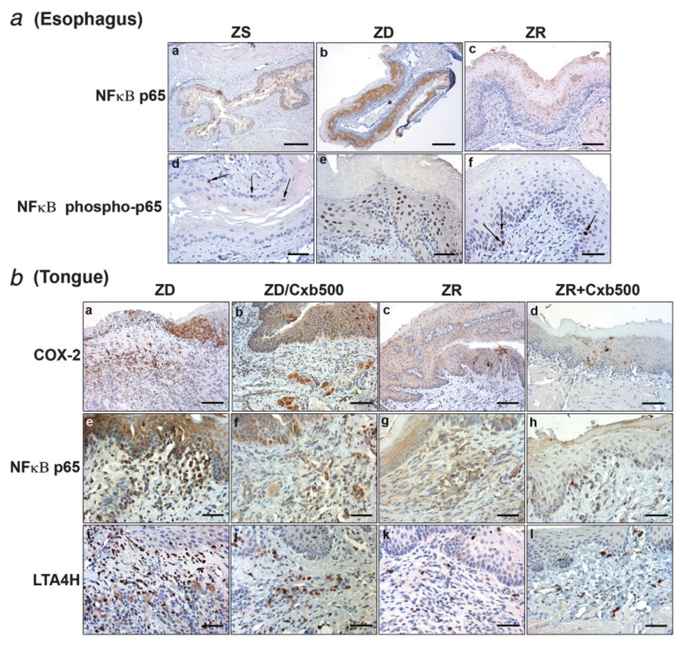
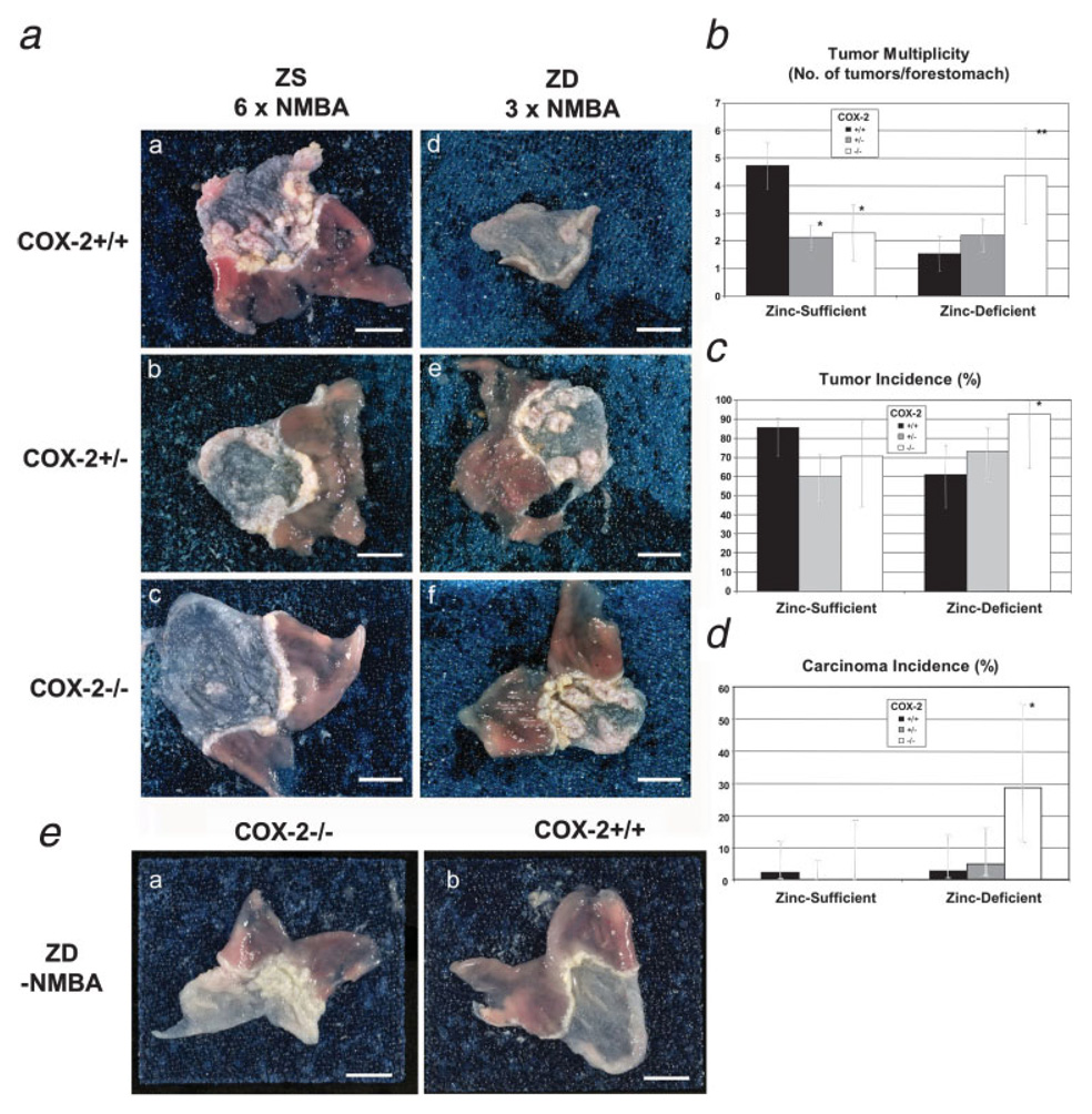
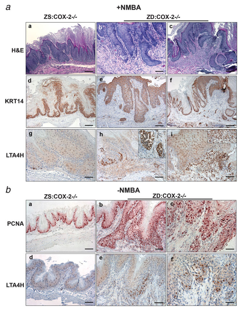
Zinc deficiency in humans is associated with an increased risk of upper aerodigestive tract (UADT) cancer. In rodents, zinc deficiency predisposes to carcinogenesis by causing proliferation and alterations in gene expression. We examined whether in zinc-deficient rodents, targeted disruption of the cyclooxygenase (COX)-2 pathway by the COX-2 selective inhibitor celecoxib or by genetic deletion prevent UADT carcinogenesis. Tongue cancer prevention studies were conducted in zinc-deficient rats previously exposed to a tongue carcinogen by celecoxib treatment with or without zinc replenishment, or by zinc replenishment alone. The ability of genetic COX-2 deletion to protect against chemically-induced forestomach tumorigenesis was examined in mice on zinc-deficient versus zinc-sufficient diet. The expression of 3 predictive biomarkers COX-2, nuclear factor (NF)-kappa B p65 and leukotriene A(4) hydrolase (LTA(4)H) was examined by immunohistochemistry. In zinc-deficient rats, celecoxib without zinc replenishment reduced lingual tumor multiplicity but not progression to malignancy. Celecoxib with zinc replenishment or zinc replenishment alone significantly lowered lingual squamous cell carcinoma incidence, as well as tumor multiplicity. Celecoxib alone reduced overexpression of the 3 biomarkers in tumors slightly, compared with intervention with zinc replenishment. Instead of being protected, zinc-deficient COX-2 null mice developed significantly greater tumor multiplicity and forestomach carcinoma incidence than wild-type controls. Additionally, zinc-deficient COX-2-/- forestomachs displayed strong LTA(4)H immunostaining, indicating activation of an alternative pathway under zinc deficiency when the COX-2 pathway is blocked. Thus, targeting only the COX-2 pathway in zinc-deficient animals did not prevent UADT carcinogenesis. Our data suggest zinc supplementation should be more thoroughly explored in human prevention clinical trials for UADT cancer.


Figures

FIGURE 1
FIGURE 1
(a) Experimental design. Weaning male rats were fed a ZS diet (40 rats) or ZD diet (229 rats) for 5 weeks and then exposed to 10 ppm NQO in deionized water for 9 weeks. NQO administration was then stopped and immediately replaced by deionized water. To determine the number of lingual lesions before the start of chemo-dietary intervention, nineteen ZD and twelve ZS rats were killed at 9 weeks. The remaining animals were taken off NQO administration. Twenty-eight ZS rats were untreated and continued on ZS diet. Two-hundred ten ZD rats were earmarked and randomly divided into 6 groups. Three groups remained on ZD diets that contained 0, 100 or 500 ppm celecoxib (Cxb), forming control ZD (48 rats), ZD/Cxb100 (24 rats) and ZD/Cxb500 (23 rats) groups. Three other groups were zinc-replenished (ZR) by switching to ZS diet, which had 0, 100 or 500 ppm Cxb, thus forming ZR (49 rats), ZR + Cxb100 (22 rats) and ZR + Cxb500 (23 rats) groups. Arrows indicate the times when animals were killed just before intervention and at 52 hr after intervention. (b) Histopathology and cell proliferation of tongues from ZS and ZD rats after exposure to NQO for 9 weeks and before intervention. Immunohistochemistry for PCNA was used to identify proliferating cells. (a, b) Hematoxylin and eosin-stained sections; (c, d) PCNA staining. ZS tongues typically showed a thickened epithelium (a) with PCNA-positive nuclei in basal and suprabasal layers (c). Six out of 19 ZD tongues showed microinvasion (b) with PCNA-positive nuclei in tumor tissue (d). (c) Histopathology of tongues at 15 weeks of intervention: (a) Invasive lingual SCC from an untreated ZD rat. (b) Invasive lingual SCC from a ZD/Cxb100 rat, (c) Invasive lingual SCC from a ZR rat and (d) Large papilloma from ZR + Cxb100 rat. Scale bars: 400 µm (c, a) 200 µm (c, b–d) and 25 µm (b, a–d).
FIGURE 2
FIGURE 2
Lingual tumor prevention by celecoxib with or without ZR in ZD rats that were formerly exposed to NQO. (a) Macroscopic appearance of tongues at 15 weeks of chemo-dietary intervention. ZD rat 53 showed multiple tumors on dorsal and lateral tongue. ZD/Cxb100 rat 61 showed a huge ulcerated tumor covering the entire posterior dorsum of the tongue. ZD/Cxb500 rat 85 had multiple large lesions on posterior dorsum and small lesions on anterior dorsum. ZR rat 111 showed 3 small lesions on the posterior dorsum of the tongue. (b–f) Tumor multiplicity, incidence of large tumors (tumors ≥ 1.5 mm), incidence of carcinoma, serum zinc levels and plasma celecoxib concentration were assessed at 15 weeks of intervention. (b) Tumor multiplicity (number of tumors per tongue). *All groups are statistically different from ZD group: ZS, ZR, ZR + Cxb100, ZR + Cxb500 and ZD/Cxb500 versus ZD, p < 0.001, ZD/Cxb100 versus ZD, p = 0.012. +Statistically different from ZD/Cxb100 group, p < 0.001. ++Statistically different from ZD/Cxb500 group, p < 0.001. (c) Large tumor incidence (%). *ZR (18 of 49), ZR+Cxb100 (7 of 22) and ZR+Cxb500 (7 of 23) versus ZD (37 of 48): p = 0.00008, p = 0.0005, p = 0.0002. ZS (8 of 28) versus ZD (24 of 48): p = 0.00007. +ZR+Cxb100 (7 of 22) versus ZD/Cxb100 (15 of 24), p = 0.045. ++ZR+Cxb500 (7 of 23) versus ZD/Cxb500 (17 of 23), p = 0.007. (d) Carcinoma incidence (%). *ZR (9 of 49), ZR+Cxb100 (3 of 22) and ZR+Cxb500 (2 of 23) versus ZD (24 of 48): p = 0.001, p = 0.004, p = 0.0006. ZS (5 of 28) versus ZD (24 of 48), p = 0.04. (e) Serum zinc levels (µg/100 mL). Sample size for each group was 10–25 rats. *Serum zinc levels in ZS, ZR, ZR+Cxb100 and ZR+Cxb500 groups are significantly greater than ZD group, p < 0.001. (f) Plasma celecoxib concentration (ng/mL). Sample size for each group was 3 rats (each from a cage with 6 animals), thus representing the celecoxib plasma content of 18 animals. All statistical tests were 2-sided. Error bars = 95% confidence intervals. Scale bars: 5 mm (a).
FIGURE 3
FIGURE 3
(a) Esophagus. Localization of NF-κB p65 and NF-κB phospho-p65 protein in archival esophageal near-serial sections from zinc-modulated rats showing COX-2 modulation by zinc (Ref. 13). (b) Tongue. COX-2, NF-κB p65 and LTA4H protein expression in tongue from NQO-treated ZD rats at 52 hr after switching to diets containing 500-ppm Cxb with ZR or without ZR. Representative sections were presented. (a) Esophagus. ZS esophagus typically showed moderately weak and sparse cytoplasmic staining of NF-κB p65 (a). NF-κB was intensely and abundantly expressed in ZD esophagi, with typical cytoplasmic and nuclear staining (b). ZR esophagus at 8 hr after zinc administration showed reduced staining (c) compared with ZD esophagus (b). Pattern of NF-κB p65 expression mirrored that of COX-2 in ZD esophagus (Ref. 13). Phospo-p65 nuclear staining was scarce and restricted to a few nuclei basal cell layer in ZS esophagus (d) but was abundant and in many cell layer in ZD esophagus (e). At 8 hr after ZR, phospho-p65 expression was sparse and occurred mostly in the nuclei of basal cells (f). (b) Tongue. At 0 hr before intervention, ZD lingual SCC showed intense COX-2 (a), NF-κB p65 (e) and LTA4H (i) protein expression in dysplastic epithelial cell layers and carcinoma. At 52 hr of intervention, ZD/Cxb500 lingual SCC showed moderately strong and frequent staining of COX-2 (b), NF-κB p65 (f) and LTA4H (j) in dysplastic epithelial cell layers and carcinoma. ZR tongues displayed diffuse cytoplasmic and isolated perinuclear staining of COX-2 (c), diffuse and moderate staining NF-κB p65 (g) and sparse occurrence of LTA4H expression in stroma (k). ZR+Cxb500 tongue had reduced and sparse occurrence of all 3 markers (COX-2, d; NF-κB p65, h and LTA4H, l). Scale bars: 100 µm (a: a and b; b: a–d); 50 µm (a: c); 25 µm (a: d–f and b: e–l).
FIGURE 4
FIGURE 4
COX-2, NF-κB p65 and LTA4H protein expression in lingual SCC at 15 weeks after switching to diets containing 100-ppm Cxb with or without ZR, or ZR alone. Representative sections were presented. Control ZD lingual SCC 47 showed strong expression of COX-2, NFκB p65 and LTA4H in carcinoma tissue and stroma (a–c). ZD/Cxb100 lingual SCC 61 had moderately strong expression of COX-2, NF-κB p65 and LTA4H in carcinoma tissue (d–f). ZR lingual SCC 123 (g–i) and ZR+Cxb100 lingual SCC 143 (j–l) showed greatly reduced COX-2 staining, absent staining of NF-κB p65 and sparse expression of LTA4H in stroma. Scale bars: 25 µm (a, b, d, e, g, h, j and k); 50 µm (c, f, i and l).
FIGURE 5
FIGURE 5
Effect of dietary zinc deficiency on NMBA-induced forestomach carcinogenesis in COX-2 deficient mice. (a) Macroscopic appearance of forestomach. ZS and ZD mice were given 6 and 3 intragastric doses of NMBA, respectively, and killed 11 weeks later. Representative sections were presented. (a–c) ZS diet. ZS:COX-2+/+ forestomach 200 had 10 tumors (a), ZS:COX-2+/− forestomach 173 showed 4 tumors (b) and ZS:COX-2−/− forestomach 199 showed 2 tumors (c). (d–f). ZD diet. ZD:COX-2+/+ forestomach 69 had 2 tumors (d), ZD:COX-2+/− forestomach 76 showed 5 tumors (e); and ZD:COX-2−/− forestomach 74 showed multiple fused tumors (f). (b) Tumor multiplicity (number of tumors per forestomach). *ZS:COX-2+/− versus ZS:COX-2+/+, p = < 0.001; ZS:COX-2−/− versus ZS:COX-2+/+, p = 0.001. **ZD:COX-2−/− versus ZD:COX-2+/+, p < 0.001. (c) Tumor incidence (%) ZD:COX-2−/− (13 of 14) versus ZD:COX-2+/+ (22 of 36), p = 0.04. (d) Carcinoma incidence (%) ZD:COX-2−/− (4 of 14) versus ZD:COX-2+/+ (1 of 36), difference = 16.3%, 95% confidence interval [CI] = 2.5–55%, p = 0.018. All statistical tests were 2-sided. (e) Macroscopic appearance of forestomach after 15 weeks of ZD diet without NMBA treatment. (a) COX−/− mouse 108 showed a thickened forestomach mucosa with lesions. (b) COX+/+ mouse 109 showed a thickened mucosa. Error bars = 95% CI. Scale bars: 5 mm (a, e).
FIGURE 6
FIGURE 6
(a) Histopathology, KRT 14 and LTA4H protein expression in zinc-sufficient ZS:COX-2−/− and zinc-deficient ZD:COX-2−/− mice forestomach at 11 weeks after NMBA treatment. (b) Effect of 15 weeks of dietary zinc deficiency in the absence of the carcinogen NMBA on cellular proliferation (assessed by PCNA immunohistochemistry) and LTA4H protein expression in COX-2−/− mice. Representative sections were presented. (a) At week 11 of NMBA treatment, ZS:COX-2−/− mouse 174 showed a thickened forestomach epithelium (a), with KRT14 expression restricted to basal and suprabasal cell layers (b) and sparse occurrence of LTA4H protein expression (c). ZD:COX-2−/− mouse 38 and 78 showed invasive forestomach SCC (b, c), with KRT14 expression in carcinoma tissue (e, f), and LTA4H expression in dysplastic and carcinoma (h, i), glandular metaplasia (inset in h) and in stroma (inset in i). (b) At week 15 of ZS or ZD diet without NMBA treatment, ZS:COX-2−/− mouse forestomach was typically thin with PCNA-positive nuclei restricted to basal cells, with weak and diffuse LTA4H protein expression in epithelium. ZD:COX-2−/− forestomach 104 and 108 showed abundant PCNA-positive cells in highly proliferative and dysplastic epithelia (b, c) and intense LTA4H protein expression in near serial forestomach sections (e, f). Scale bars: 100 µm (a: a, c, f); 50 µm (a: b, d, e, g–i; b: a–c) and 25 µm (b: d, c, f).

Similar articles

Cited by

References

    1. Parkin DM, Bray F, Ferlay J, Pisani P. Global cancer statistics, 2002. CA Cancer J Clin. 2005;55:74–108. - PubMed
    1. Moore SR, Johnson NW, Pierce AM, Wilson DF. The epidemiology of tongue cancer: a review of global incidence. Oral Dis. 2000;6:75–84. - PubMed
    1. Makuuchi H, Machimura T, Shimada H, Mizutani K, Chino O, Kise Y, Nishi T, Tanaka H, Mitomi T, Horiuchi M, Sakai M, Gotoh J, et al. Endoscopic screening for esophageal cancer in 788 patients with head and neck cancers. Tokai J Exp Clin Med. 1996;21:139–145. - PubMed
    1. Slaughter DP, Southwick HW, Smejkal W. Field cancerization in oral stratified squamous epithelium; clinical implications of multicentric origin. Cancer. 1953;6:963–968. - PubMed
    1. Doerr TD, Prasad AS, Marks SC, Beck FW, Shamsa FH, Penny HS, Mathog RH. Zinc deficiency in head and neck cancer patients. J Am Coll Nutr. 1997;16:418–422. - PubMed

Publication types

MeSH terms