. 2007 Nov 28;129(47):14746-51.
doi: 10.1021/ja0754002. Epub 2007 Nov 7.

Biophysical and structural characterization of a robust octameric beta-peptide bundle

Affiliations

Biophysical and structural characterization of a robust octameric beta-peptide bundle

Jessica L Goodman et al. J Am Chem Soc. .

Abstract

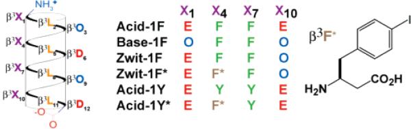
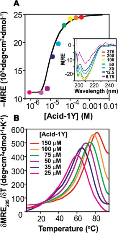
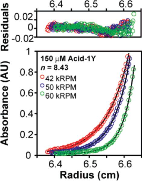
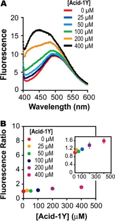
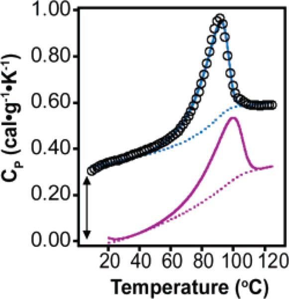
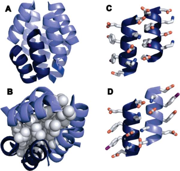
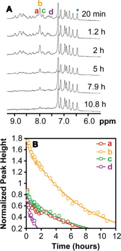
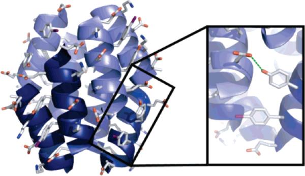
Proteins composed of alpha-amino acids are essential components of the machinery required for life. Stanley Miller's renowned electric discharge experiment provided evidence that an environment of methane, ammonia, water, and hydrogen was sufficient to produce alpha-amino acids. This reaction also generated other potential protein building blocks such as the beta-amino acid beta-glycine (also known as beta-alanine); however, the potential of these species to form complex ordered structures that support functional roles has not been widely investigated. In this report we apply a variety of biophysical techniques, including circular dichroism, differential scanning calorimetry, analytical ultracentrifugation, NMR and X-ray crystallography, to characterize the oligomerization of two 12-mer beta3-peptides, Acid-1Y and Acid-1Y*. Like the previously reported beta3-peptide Zwit-1F, Acid-1Y and Acid-1Y* fold spontaneously into discrete, octameric quaternary structures that we refer to as beta-peptide bundles. Surprisingly, the Acid-1Y octamer is more stable than the analogous Zwit-1F octamer, in terms of both its thermodynamics and kinetics of unfolding. The structure of Acid-1Y, reported here to 2.3 A resolution, provides intriguing hypotheses for the increase in stability. To summarize, in this work we provide additional evidence that nonnatural beta-peptide oligomers can assemble into cooperatively folded structures with potential application in enzyme design, and as medical tools and nanomaterials. Furthermore, these studies suggest that nature's selection of alpha-amino acid precursors was not based solely on their ability to assemble into stable oligomeric structures.


Figures

Figure 1
Figure 1
Helical net representation of bundle-forming β3-peptides and the structure of the β3-4-iodohomophenylalanine residue found in Zwit-1F* and Acid-1Y*. Previously characterized β-peptide bundles include Zwit-1F and Zwit-1F*– as well as a 1:1 mixture of Acid-1F and Base-1F (D. Daniels, J. Qiu, A. Schepartz, manuscript in preparation).
Figure 2
Figure 2
Acid-1Y self-association monitored by circular dichroism spectroscopy (CD). (A) Plot of MRE208 as a function of [Acid-1Y]T fit to a monomer–octamer equilibrium. (Inset) Wavelength-dependent CD spectra of Acid-1Y (MRE208 in units of 103 deg·cm2·dmol–1) at the indicated [Acid-1Y]T (μM). (B) Plot of δMRE205·δT–1 for the concentrations of Acid-1Y shown.
Figure 3
Figure 3
Acid-1Y self-association monitored by analytical ultracentrifugation (AU) (150 μM Acid-1Y) and fit to monomer-n-mer equilibria where n = 8.4. Samples were prepared in 10 mM NaH2PO4, 200 mM NaCl (pH 7.1) and centrifuged to equilibrium at 25 °C at speeds of 42 (red), 50 (green), or 60 (blue) kRPM. The experimental data points are shown as open circles; lines indicate a fit to a monomer–octamer model as described in the experimental section (see Supporting Information).
Figure 4
Figure 4
Fluorescence of 1-anilino-8-naphthalenesulfonate (ANS) in the presence and absence of Acid-1Y. (A) Change in fluorescence of 10 μM ANS in the presence of the indicated concentration of Acid-1Y. Binding reactions were prepared in a buffer composed of 10 × PBC (100 mM phosphate, borate, and citrate) and 200 mM NaCl (pH 7.0). (B). Ratio of ANS fluorescence in the presence of given concentrations of Acid-1Y relative to fluorescence in the absence of peptide (10 μM ANS) as calculated using the global maximum fluorescence value for each concentration (observed at 492, 497, 491, 490, 487, 433 nm for concentrations of 0, 25, 50, 100, 200, and 400 μM Acid-1Y, respectively). (Inset) Fluorescence ratio of Acid-1Y as a function of concentration on a smaller scale to show the error bars.
Figure 5
Figure 5
DSC analysis of 300 μM Acid-1Y fit to a subunit dissociation model as described in Supporting Information. Raw data are shown as black circles, the calculated thermogram is shown as a blue line, and the baseline as a dotted blue line. The calculated thermogram and baseline for 300 μM Zwit-1F are shown as purple solid and dotted lines, respectively. The Acid-1Y data set is offset artificially for clarity, as indicated by the arrow.
Figure 6
Figure 6
Crystal structure of Acid-1Y*. (A) Ribbon diagram of Acid-1Y* octamer with parallel helices represented by similar shading. The rmsd is 20 Å between the previously solved Zwit-1F and the current Acid-1Y structures. (B) Space-filling model of leucine side-chain packing in the Acid-1Y* core. Ribbon diagram of (C) parallel and (D) antiparallel helices illustrating side-chain interactions.
Figure 7
Figure 7
Hydrogen/deuterium NMR exchange analysis of the Acid-1Y octamer. (A) 500 MHz 1H NMR spectra of 0.750 mM Acid-1Y acquired at the indicated times after a lyophilized Acid-1Y sample was reconstituted in phosphate-buffered D2O. (B) The normalized heights of the indicated resonances (normalized to the aromatic resonance at 6.39 ppm (*)) fit to exponential function.
Figure 8
Figure 8
Structure of Acid-1Y with tyrosine side chains rendered as sticks illustrates hydrogen-bond formation between a tyrosine residue of one helix with an aspartic acid residue from another helix, showing how the tyrosine hydroxyl could provide additional stability through hydrogen-bond formation.

References

    1. Svedberg T, Fahraeus R. J. Am. Chem. Soc. 1926;48:430–438.
    1. Klotz IM, Langerman NR, Darnall DW. Annu. Rev. Biochem. 1970;39:25–62. - PubMed
    1. Shimojima A, Liu Z, Ohsuna T, Terasaki O, Kuroda K. J. Am. Chem. Soc. 2005;127:14108–14116. - PubMed
    1. Mirkin CA, Letsinger RL, Mucic RC, Storhoff JJ. Nature. 1996;382:607–609. - PubMed
    1. Hartgerink JD, Clark TD, Ghadiri RM. Chem. Eur. J. 1998;4:1367–1372.

Publication types