

Inherent antibacterial activity of a peptide-based beta-hairpin hydrogel
- PMID: 17985907
- PMCID: PMC2650250
- DOI: 10.1021/ja076300z
Inherent antibacterial activity of a peptide-based beta-hairpin hydrogel
Abstract
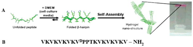
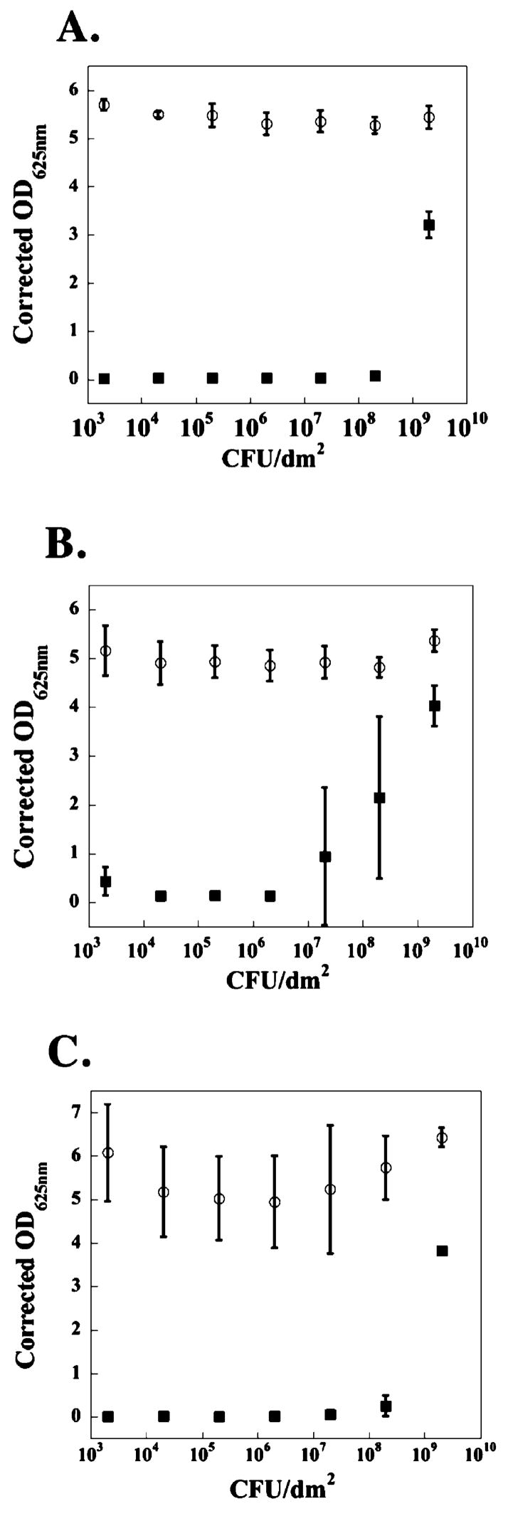
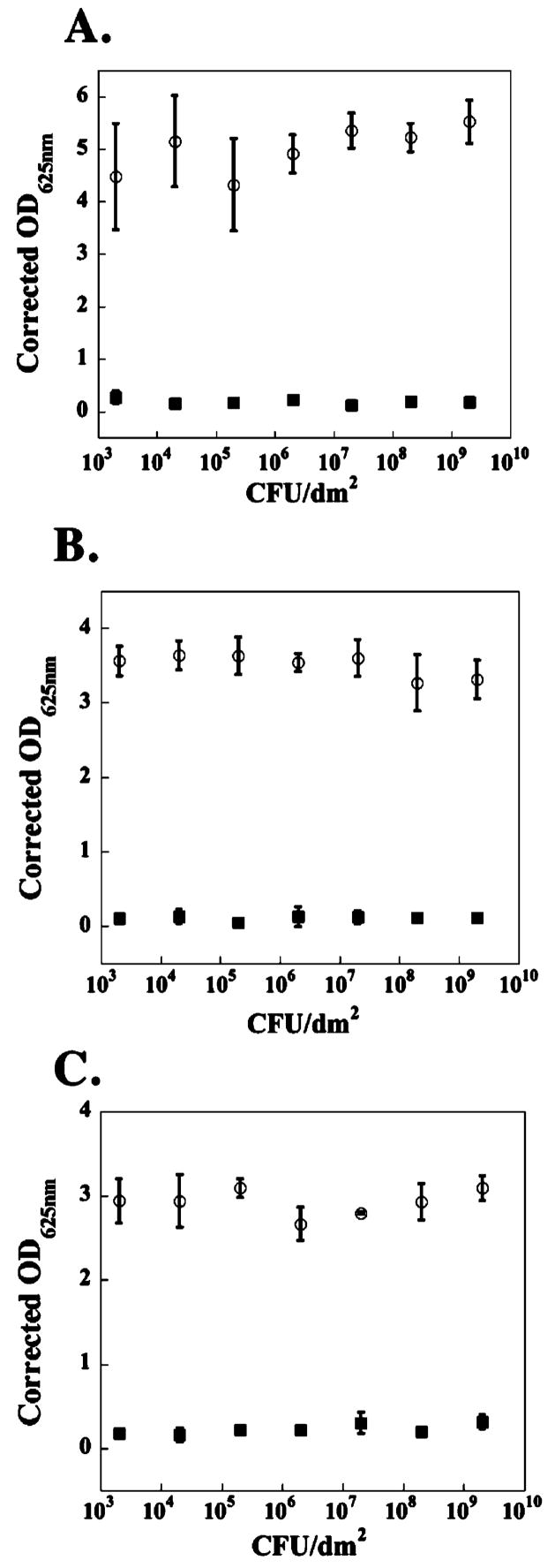
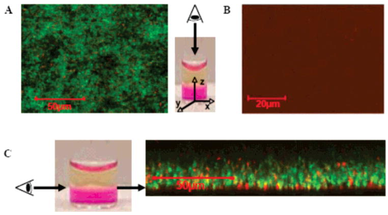
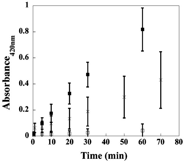
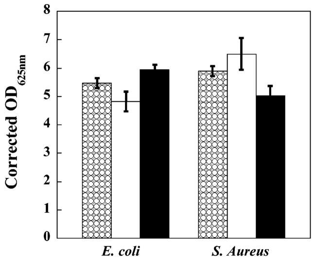
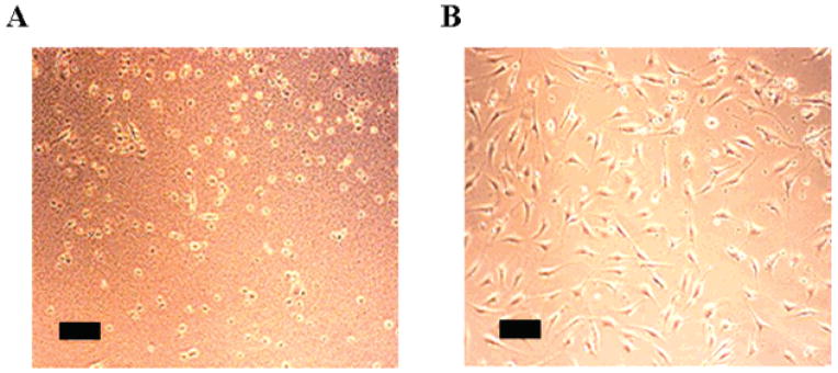
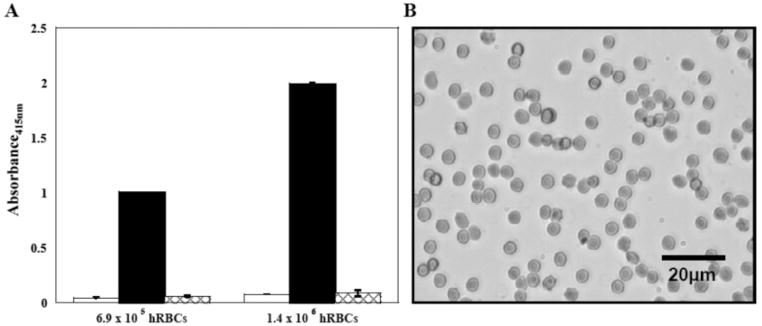
Among several important considerations for implantation of a biomaterial, a main concern is the introduction of infection. We have designed a hydrogel scaffold from the self-assembling peptide, MAX1, for tissue regeneration applications whose surface exhibits inherent antibacterial activity. In experiments where MAX1 gels are challenged with bacterial solutions ranging in concentrations from 2 x 10(3) colony forming units (CFUs)/dm2 to 2 x 10(9) CFUs/dm2, gel surfaces exhibit broad-spectrum antibacterial activity. Results show that the hydrogel surface is active against Gram-positive (Staphylococcus epidermidis, Staphylococcus aureus, and Streptococcus pyogenes) and Gram-negative (Klebsiella pneumoniae and Escherichia coli) bacteria, all prevalent in hospital settings. Live-dead assays employing laser scanning confocal microscopy show that bacteria are killed when they engage the surface. In addition, the surface of MAX1 hydrogels was shown to cause inner and outer membrane disruption in experiments that monitor the release of beta-galactosidase from the cytoplasm of lactose permease-deficient E. coli ML-35. These data suggest a mechanism of antibacterial action that involves membrane disruption that leads to cell death upon cellular contact with the gel surface. Although the hydrogel surface exhibits bactericidal activity, co-culture experiments indicate hydrogel surfaces show selective toxicity to bacterial versus mammalian cells. Additionally, gel surfaces are nonhemolytic toward human erythrocytes, which maintain healthy morphologies when in contact with the surface. These material attributes make MAX1 gels attractive candidates for use in tissue regeneration, even in nonsterile environments.
Figures
References
-
-
For recent reviews, see refs –: Drury JL, Mooney DJ. Biomaterials. 2003;24:4337–4351.
-
-
- Fairman R, Akerfeldt KS. Curr Opin Struct Biol. 2005;15:453–463. - PubMed
-
- Hoffman AS. Adv Drug Delivery Rev. 2002;54:3–12. - PubMed
-
- Lutolf MP, Hubbell JA. Nat Biotechnol. 2005;23:47–55. - PubMed
-
- Mart RJ, Osborne RD, Stevens MM, Ulijn RV. Soft Matter. 2006;2:822–835. - PubMed
Publication types
MeSH terms
Substances
Grants and funding
LinkOut - more resources
Full Text Sources
Other Literature Sources
Medical
