

In vitro selection of histone H4 aptamers for recognition imaging microscopy
- PMID: 17985909
- PMCID: PMC2533041
- DOI: 10.1021/ja076488m
In vitro selection of histone H4 aptamers for recognition imaging microscopy
Abstract
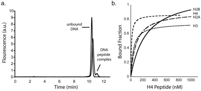
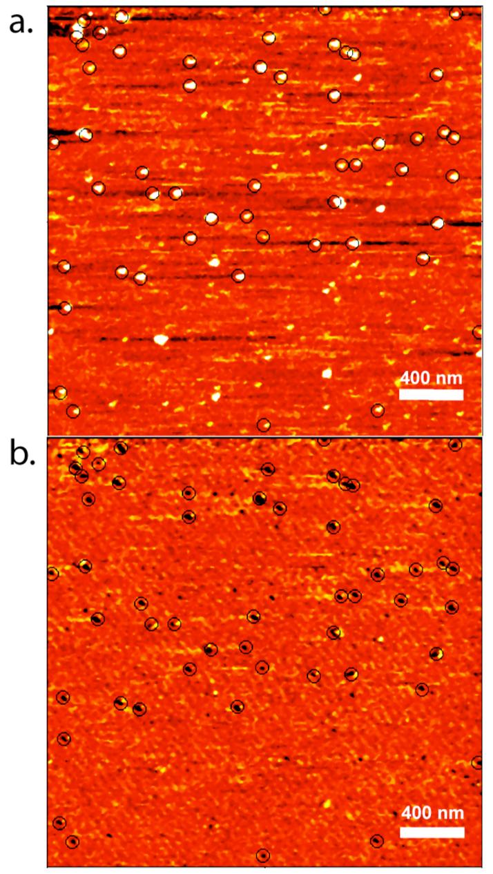
Recognition imaging microscopy is an analytical technique used to map the topography and chemical identity of specific protein molecules present in complex biological samples. The technique relies on the use of antibodies tethered to the cantilever tip of an AFM probe to detect cognate antigens deposited onto a mica surface. Despite the power of this technique to resolve single molecules with nanometer-scale spacing, the recognition step remains limited by the availability of suitable quality antibodies. Here we report the in vitro selection and recognition imaging of anti-histone H4 aptamers. In addition to identifying aptamers to highly basic proteins, these results suggest that aptamers provide an efficient, cost-effective route to highly selective affinity reagents for recognition imaging microscopy.
Figures
References
-
- Kienberger F, Ebner A, Bruber HJ, Hinterdorfer P. Acc. Chem. Res. 2006;39:29–36. - PubMed
-
- Raab A, Han W, Badt D, Smith-Gill SJ, Lindsay SM, Schindler H, Hinterdorfer P. Nature Biotech. 1999;17:902–905. - PubMed
-
- Bash R, Wang H, Anderson C, Yodh J, Hager G, Lindsay SM, Lohr D. FEBS Lett. 2006;580:4757–4761. - PubMed
Publication types
MeSH terms
Substances
Grants and funding
LinkOut - more resources
Full Text Sources
Other Literature Sources
Miscellaneous
