

Maternal heparin-binding-EGF deficiency limits pregnancy success in mice
- PMID: 17986609
- PMCID: PMC2084340
- DOI: 10.1073/pnas.0707909104
Maternal heparin-binding-EGF deficiency limits pregnancy success in mice
Abstract
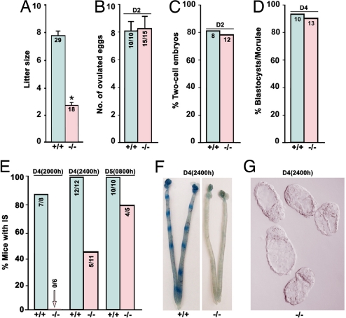
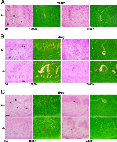
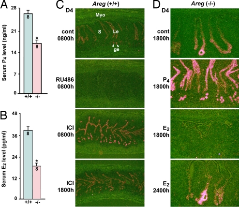
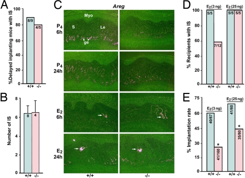
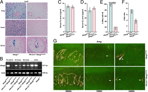
An intimate discourse between the blastocyst and uterus is essential for successful implantation. However, the molecular basis of this interaction is not clearly understood. Exploiting genomic Hbegf mutant mice, we show here that maternal deficiency of heparin-binding EGF-like growth factor (HB-EGF) defers on-time implantation, leading to compromised pregnancy outcome. We also demonstrate that amphiregulin, but not epiregulin, partially compensates for the loss of HB-EGF during implantation. In search of the mechanism of this compensation, we found that reduced preimplantation estrogen secretion from ovarian HB-EGF deficiency is a cause of sustained expression of uterine amphiregulin before the initiation of implantation. To explore the significance specifically of uterine HB-EGF in implantation, we examined this event in mice with conditional deletion of uterine HB-EGF and found that this specific loss of HB-EGF in the uterus still defers on-time implantation without altering preimplantation ovarian estrogen secretion. The observation of normal induction of uterine amphiregulin surrounding the blastocyst at the time of attachment in these conditional mutant mice suggests a compensatory role of amphiregulin for uterine loss of HB-EGF, preventing complete failure of pregnancy. Our study provides genetic evidence that HB-EGF is critical for normal implantation. This finding has high clinical relevance, because HB-EGF signaling is known to be important for human implantation.
Conflict of interest statement
The authors declare no conflict of interest.
Figures
Similar articles
-
Dual source and target of heparin-binding EGF-like growth factor during the onset of implantation in the hamster.Development. 2002 Sep;129(17):4125-34. doi: 10.1242/dev.129.17.4125. Development. 2002. PMID: 12163414
-
Dysregulation of EGF family of growth factors and COX-2 in the uterus during the preattachment and attachment reactions of the blastocyst with the luminal epithelium correlates with implantation failure in LIF-deficient mice.Mol Endocrinol. 2000 Aug;14(8):1147-61. doi: 10.1210/mend.14.8.0498. Mol Endocrinol. 2000. PMID: 10935540
-
Heparin-binding EGF-like growth factor gene is induced in the mouse uterus temporally by the blastocyst solely at the site of its apposition: a possible ligand for interaction with blastocyst EGF-receptor in implantation.Development. 1994 May;120(5):1071-83. doi: 10.1242/dev.120.5.1071. Development. 1994. PMID: 8026321
-
HB-EGF: a unique mediator of embryo-uterine interactions during implantation.Exp Cell Res. 2009 Feb 15;315(4):619-26. doi: 10.1016/j.yexcr.2008.07.025. Epub 2008 Aug 3. Exp Cell Res. 2009. PMID: 18708050 Free PMC article. Review.
-
Diverse functions of HBEGF during pregnancy.Mol Reprod Dev. 2009 Dec;76(12):1116-27. doi: 10.1002/mrd.21066. Mol Reprod Dev. 2009. PMID: 19565643 Free PMC article. Review.
Cited by
-
Minireview: Steroid-regulated paracrine mechanisms controlling implantation.Mol Endocrinol. 2014 Sep;28(9):1408-22. doi: 10.1210/me.2014-1074. Epub 2014 Jul 22. Mol Endocrinol. 2014. PMID: 25051170 Free PMC article. Review.
-
Nuclear Shp2 directs normal embryo implantation via facilitating the ERα tyrosine phosphorylation by the Src kinase.Proc Natl Acad Sci U S A. 2017 May 2;114(18):4816-4821. doi: 10.1073/pnas.1700978114. Epub 2017 Apr 19. Proc Natl Acad Sci U S A. 2017. PMID: 28424251 Free PMC article.
-
In vitro effects of heparin-binding epidermal growth factor on adhesion stage of implantation.Rom J Morphol Embryol. 2023 Oct-Dec;64(4):493-500. doi: 10.47162/RJME.64.4.05. Rom J Morphol Embryol. 2023. PMID: 38184829 Free PMC article.
-
Molecular Signaling Regulating Endometrium-Blastocyst Crosstalk.Int J Mol Sci. 2019 Dec 18;21(1):23. doi: 10.3390/ijms21010023. Int J Mol Sci. 2019. PMID: 31861484 Free PMC article. Review.
-
Restraint stress inhibits mouse implantation: temporal window and the involvement of HB-EGF, estrogen and progesterone.PLoS One. 2013 Nov 14;8(11):e80472. doi: 10.1371/journal.pone.0080472. eCollection 2013. PLoS One. 2013. PMID: 24244689 Free PMC article.
References
-
- Wang H, Dey SK. Nat Rev Genet. 2006;7:185–199. - PubMed
-
- Das SK, Das N, Wang J, Lim H, Schryver B, Plowman GD, Dey SK. Dev Biol. 1997;190:178–190. - PubMed
-
- Tamada H, Das SK, Andrews GK, Dey SK. Biol Reprod. 1991;45:365–372. - PubMed
-
- Das SK, Chakraborty I, Paria BC, Wang XN, Plowman G, Dey SK. Mol Endocrinol. 1995;9:691–705. - PubMed
-
- Das SK, Wang XN, Paria BC, Damm D, Abraham JA, Klagsbrun M, Andrews GK, Dey SK. Development (Cambridge, UK) 1994;120:1071–1083. - PubMed
Publication types
MeSH terms
Substances
Grants and funding
- R03 HD050315/HD/NICHD NIH HHS/United States
- HD12304/HD/NICHD NIH HHS/United States
- F31 DA021062/DA/NIDA NIH HHS/United States
- R01 HD037830/HD/NICHD NIH HHS/United States
- U54 HD033994/HD/NICHD NIH HHS/United States
- HD33994/HD/NICHD NIH HHS/United States
- P30 HD033994/HD/NICHD NIH HHS/United States
- DA06668/DA/NIDA NIH HHS/United States
- R37 HD012304/HD/NICHD NIH HHS/United States
- HD37830/HD/NICHD NIH HHS/United States
- HD50315/HD/NICHD NIH HHS/United States
- U54 HD028934/HD/NICHD NIH HHS/United States
- U54-HD28934/HD/NICHD NIH HHS/United States
- R01 DA006668/DA/NIDA NIH HHS/United States
- ES07814/ES/NIEHS NIH HHS/United States
- R01 ES007814/ES/NIEHS NIH HHS/United States
- R37 DA006668/DA/NIDA NIH HHS/United States
LinkOut - more resources
Full Text Sources
Molecular Biology Databases
