. 2007 Nov 13;104(46):17995-8000.
doi: 10.1073/pnas.0705038104. Epub 2007 Nov 6.

Folding of noncoding RNAs during transcription facilitated by pausing-induced nonnative structures

Affiliations

Folding of noncoding RNAs during transcription facilitated by pausing-induced nonnative structures

Terrence N Wong et al. Proc Natl Acad Sci U S A. .

Abstract

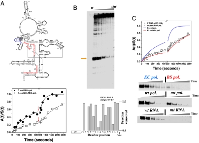
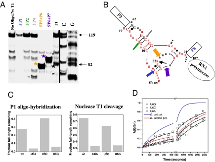
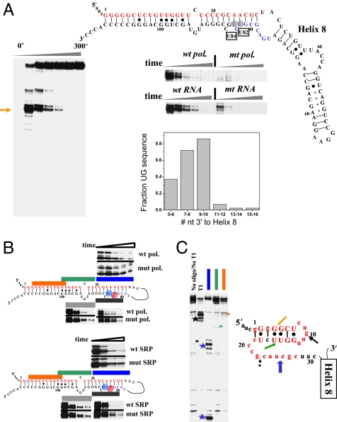
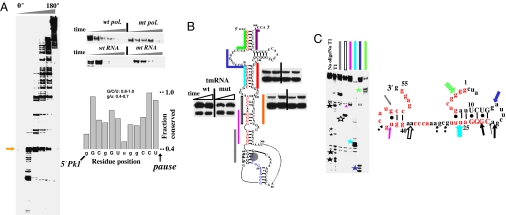
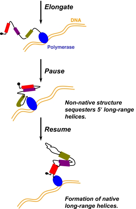
RNA folding in the cell occurs during transcription. Expedient RNA folding must avoid the formation of undesirable structures as the nascent RNA emerges from the RNA polymerase. We show that efficient folding during transcription of three conserved noncoding RNAs from Escherichia coli, RNase P RNA, signal-recognition particle RNA, and tmRNA is facilitated by their cognate polymerase pausing at specific locations. These pause sites are located between the upstream and downstream portions of all of the native long-range helices in these noncoding RNAs. In the paused complexes, the nascent RNAs form labile structures that sequester these upstream portions in a manner to possibly guide folding. Both the pause sites and the secondary structure of the nonnative portions of the paused complexes are phylogenetically conserved among gamma-proteobacteria. We propose that specific pausing-induced structural formation is a general strategy to facilitate the folding of long-range helices. This polymerase-based mechanism may result in portions of noncoding RNA sequences being evolutionarily conserved for efficient folding during transcription.


Conflict of interest statement

The authors declare no conflict of interest.

Figures

Fig. 1.
Fig. 1.
Folding and pausing of E. coli P RNA during transcription. (A) At the top, E. coli P RNA. Upstream regions of six long-range helices before the 119 pause (shaded) are in red. At the bottom, folding of P RNA when transcribed by E. coli polymerase [k1 = (15 ± 4) × 10−3·s−1 and f1 = 0.23; k2 = (3 ± 1) × 10−3·s−1 and f2 = 0.77] or by B. subtilis polymerase [k1 = (13 ± 4) × 10−3·s−1 and f1 = 0.21; k2 = (0.5 ± 0.1) × 10−3·s−1 and f2 = 0.79]. Red arrows: +rifampicin to halt transcription. (B Upper) The C119 pause site (orange arrow). (Lower) Fifteen nucleotides preceding the 119 pause manually aligned to show conservation among γ-proteobacteria (53 sequences). (C Upper) The effect on P RNA folding during transcription by the mutant E. coli polymerase [k1 = (12 ± 1)×10−3·s−1 and f1 = 0.30; k2 = (0.5 ± 0.1) ×10−3·s−1 and f2 = 0.70] or by the wild-type polymerase of the C119→G/G107→C mutant [k1 = (10 ± 1)×10−3·s−1 and f1 = 0.33; k2 = (0.5 ± 0.1)×10−3·s−1 and f2 = 0.67]. (Lower) Comparisons of 119 pause during transcription by E. coli and B. subtilis polymerase by wild-type and the mutant E. coli polymerase, and by wild-type polymerase by using the C119→G/G107→C mutant template.
Fig. 2.
Fig. 2.
Structural analysis of the nascent RNA in the 119-paused complex. (A) Structural mapping of 5′ 32P-labeled 119-paused complex by oligohybridization by using probes complementary to different regions in P RNA and by partial nuclease T1 digestion. The cleavage products are indicated by stars. (B) Proposed structure of the nascent RNA in the 119-paused complex. Colored block arrows show the approximate location and relative intensity of RNase H cleavage upon oligohybridization with the color matching the oligo probe used in A. A black arrow shows the location of the major T1 cleavage product (large star in A). Minor T1 cleavage products are dashed (small stars in A). Red nucleotides indicate the 5′ nucleotides of the long-range helices P1, P2, P4, P5, P6, and P7. Phylogenetically supported base pairs are shown in capital letters. (C) Structural mapping of U6 mutants by T1 nuclease cleavage and by P1 oligohybridization. Mapping with the P2, P4, P5-P6, and P6-P7 oligoprobes produced results similar to wild type (not shown). (D) Folding of U6 mutants when transcribed by E. coli polymerase.
Fig. 3.
Fig. 3.
Pausing, folding, and structural analysis of E. coli SRP RNA. (A Left) Two pause sites (U82 and U84) are present during transcription by wild-type polymerase. (Right) These pause sites are greatly reduced in the transcription by the mutant polymerase, or upon mutating U82 and its base pairing partner A25 to A82-U25. The graph shows the presence of 5′ UG 7–10 nucleotides downstream to the native helix-8 (manual alignment of 43 sequences). Upstream portions of long-range helices are in red, and the U82/U84 pauses are boxed. (B) Folding of SRP RNA monitored by oligohybridization. (Upper) Transcription by wild-type and mutant polymerase. (Lower) Transcription of wild-type and U25-A82 mutant. The time points are from 15–300 seconds. (C Left) Structural mapping of the body-labeled U82/U84 paused complex by oligohybridization and partial nuclease T1 digestion. The three oligoprobes are complementary to the upstream portions of long-range helices (matching colors as those in B). The cleavage products are indicated by stars. (Right) Proposed structure of the nascent RNA in paused complex. Phylogenetically supported base pairs are shown in capital letters. Colored block arrows, relative intensity and location of RNase H/oligohybridization with the color matching the oligo probe. Black arrows, location of major T1 cleavage (large star in gel on the left) with dashed arrows indicating minor cleavage product (small star in gel on the left).
Fig. 4.
Fig. 4.
Pausing, folding, and structural analysis of E. coli tmRNA. (A Left and Upper Right) Many pause sites are present during transcription by wild-type polymerase. The first major pause (arrow) at U65 is less pronounced in the transcription by the mutant polymerase or through mutation of A51-U65 to U51-A65. Many pause sites are present in the pseudoknot region (residues 200–280). (Lower Right) Sequence preceding the U65 pause is conserved among γ-proteobacteria (manual alignment of 69 sequences). Nucleotides exhibiting especially high levels of conservation are capitalized. (B) Folding monitored by oligohybridization during transcription of wild-type and U25-A82 pausing deficient tmRNA by wild-type polymerase. Upstream portions of the long-range helices are in red and the U65 pause site is shaded. The relative protection from 15–30 seconds is shown. (C Left) Structural mapping of the body-labeled U65 paused complex by oligohybridization and partial nuclease T1 digestion. The six oligos used are complementary to the upstream portions of the long-range helices (matching colors as those in B). The cleavage products are indicated by stars. (Right) Proposed structure of the nascent RNA in paused complex. Phylogenetically supported base pairs are shown in capital letters. Colored block arrows, relative intensity and location of RNase H/oligohybridization with color matching the oligo probe. Black arrows, location of the major T1 cleavage (large stars in gel on the left) with dashed arrows indicating minor cleavage products (small stars in gels on the left).
Fig. 5.
Fig. 5.
Pausing, long-range helices, and nonnative structure during folding during transcription. DNA template is in gold, and RNA polymerase is in blue. RNA residues involved in long-range helices are shown as filled bars (different colors represent different helices), and other regions of the nascent RNA transcript are shown as black lines. Interaction between regions in the RNA transcript is indicated by proximity.

References

    1. Treiber DK, Williamson JR. Curr Opin Struct Biol. 2001;11:309–314. - PubMed
    1. Woodson SA. Biochem Soc Trans. 2002;30:1166–1169. - PubMed
    1. Sosnick TR, Pan T. Curr Opin Struct Biol. 2003;13:309–316. - PubMed
    1. Isambert H, Siggia ED. Proc Natl Acad Sci USA. 2000;97:6515–6520. - PMC - PubMed
    1. Woodson SA. Cell Mol Life Sci. 2000;57:796–808. - PMC - PubMed

Publication types

LinkOut - more resources