. 2007 Nov 7;2(11):e1150.
doi: 10.1371/journal.pone.0001150.

Role of matrix metalloproteinase 13 in both endochondral and intramembranous ossification during skeletal regeneration

Affiliations

Role of matrix metalloproteinase 13 in both endochondral and intramembranous ossification during skeletal regeneration

Danielle J Behonick et al. PLoS One. .

Abstract

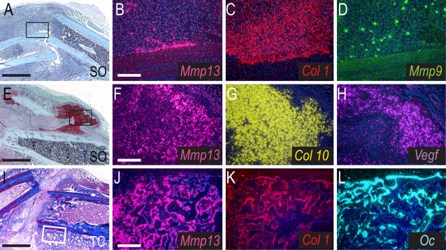
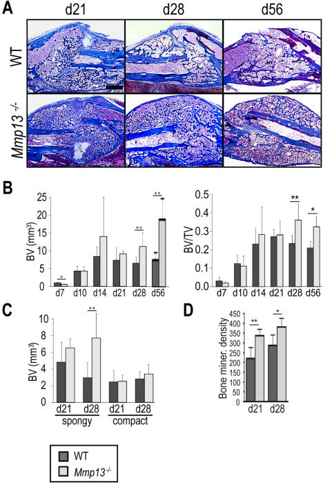
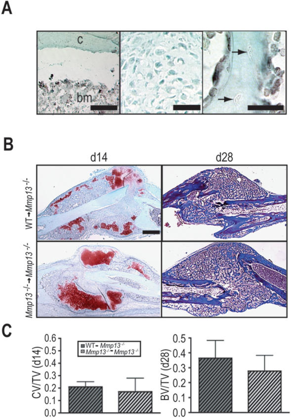
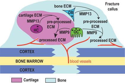
Extracellular matrix (ECM) remodeling is important during bone development and repair. Because matrix metalloproteinase 13 (MMP13, collagenase-3) plays a role in long bone development, we have examined its role during adult skeletal repair. In this study we find that MMP13 is expressed by hypertrophic chondrocytes and osteoblasts in the fracture callus. We demonstrate that MMP13 is required for proper resorption of hypertrophic cartilage and for normal bone remodeling during non-stabilized fracture healing, which occurs via endochondral ossification. However, no difference in callus strength was detected in the absence of MMP13. Transplant of wild-type bone marrow, which reconstitutes cells only of the hematopoietic lineage, did not rescue the endochondral repair defect, indicating that impaired healing in Mmp13-/- mice is intrinsic to cartilage and bone. Mmp13-/- mice also exhibited altered bone remodeling during healing of stabilized fractures and cortical defects via intramembranous ossification. This indicates that the bone phenotype occurs independently from the cartilage phenotype. Taken together, our findings demonstrate that MMP13 is involved in normal remodeling of bone and cartilage during adult skeletal repair, and that MMP13 may act directly in the initial stages of ECM degradation in these tissues prior to invasion of blood vessels and osteoclasts.


Conflict of interest statement

Competing Interests: The authors have declared that no competing interests exist.

Figures

Figure 1
Figure 1. Mmp13 expression during non-stabilized fracture healing.
(Left column) Safranin-O/Fast Green (SO) and Trichrome (TC) stained sagittal sections through the WT callus at 6 (A), 10 (E) and 14 (I) days post-fracture. Cartilage (red) develops during the soft callus phase of healing (A), is resorbed during the hard callus phase (E) and is replaced by bone (blue, I). (Middle/Right column) In situ hybridization analyses of Mmp13 expression and osteoblast/chondrocyte differentiation markers. (B–D) At 6 days post-fracture, Mmp13 is expressed in the callus and overlaps with Col1-expressing cells (early osteoblasts) but not Mmp9-expressing cells (osteoclasts and inflammatory cells). (F–H) At day 10, Mmp13 mRNA is detected in hypertrophic chondrocytes also expressing Col10 and Vegf. (J–L) At day 14, Mmp13 is expressed in mature osteoblasts co-expressing Col1 and/or Oc. Scale bars: A, E, I = 1mm; B-D, F-H, J-L-200 µm.
Figure 2
Figure 2. Mmp13 −/− mice display an accumulation of cartilage during non-stabilized fracture healing.
(A) SO staining of WT and Mmp13 −/− fracture callus at 7, 10, 14, and 21 days post-fracture shows that cartilage persists in the Mmp13 −/− callus from 14 through 21 days post-fracture. Scale bar = 1 mm (B) Histomorphometric measurements of total callus volume (TV), total cartilage volume (CV) and total cartilage volume as a proportion of total callus volume (CV/TV) in WT and Mmp13 −/− mice at day 7 (WT n = 6, Mmp13 −/− n = 6), 10 (WT n = 8, Mmp13 −/− n = 6), 14 (WT n = 8, Mmp13 −/− n = 6), 21 (WT n = 6, Mmp13 −/− n = 6) and 28 (WT n = 6, Mmp13 −/− n = 6). There is a statistically significant increase in total cartilage volume in Mmp13 −/− calluses compared with WT at day 7 (**p<0.01), 14 (*p<0.05) and 21 (*p<0.05). There is a statistically significant increase in total cartilage volume as a proportion of total callus volume in Mmp13 −/− calluses compared with WT at day 7 (**p<0.01), 14 (*p<0.05) and 21 (*p<0.05). Wilcoxon test, bars represent means ± S.D. At 21 days post-fracture, all Mmp13−/− calluses contained cartilage as compared to 1/3 of WT. At 28 days post-fracture, only Mmp13−/− calluses (1/3) still contained cartilage.
Figure 3
Figure 3. Integral steps in endochondral ossification are unperturbed during non-stabilized fracture repair in Mmp13−/− mice.
(A) Overlay of SO stained sections with in situ hybridization for Col2 (red) indicate no difference in the early differentiation of chondrocytes in WT and Mmp13 −/− calluses at day 5. Overlay of SO stained sections with in situ hybridization for Col10 (yellow) shows a delay in hypertrophic chondrocyte removal in the Mmp13 −/− callus at day 14. Scale bar = 1 mm (B) Cellular analyses of WT and Mmp13 −/− calluses at day 14 show that blood vessels (PECAM) and osteoclasts (TRAP) are present in the Mmp13 −/− callus while aggrecan cleavage by MMPs (DIPEN epitope) is reduced in the Mmp13 −/− callus. Scale bar = 1 mm
Figure 4
Figure 4. Mmp13 −/− mice display increased bone volume during non-stabilized fracture healing.
(A) Trichrome staining of WT and Mmp13 −/− non-stabilized fracture calluses shows an increase in the amount of bone in the Mmp13 −/− callus compared to WT at days 28 and 56 post-fracture. Scale bar = 1 mm (B) Histomorphometric measurements of total bone volume (BV) and total bone volume as a proportion of total callus volume (BV/TV) in WT and Mmp13 −/− mice at days 7 (WT n = 6, Mmp13 −/− n = 6), 10 (WT n = 8, Mmp13 −/− n = 6), 14 (WT n = 8, Mmp13 −/− n = 6), 21 (WT n = 6, Mmp13 −/− n = 6), 28 (WT n = 6, Mmp13 −/− n = 6) and 56 (WT n = 5, Mmp13 −/− n = 4) confirm this observation There is a statistically significant decrease in BV in Mmp13 −/− calluses compared with WT at day 7 (*p<0.05), but a statistically significant increase in BV and BV/TV in Mmp13 −/− calluses compared with WT at days 28 (**p<0.01 and p<0.05 respectively) and 56 (*p<0.05). Wilcoxon test, bars represent means ± S.D. (C) Histomorphometric measurements indicate a statistically significant difference in total spongy bone volume in Mmp13 −/− calluses compared with WT at day 28 (**p<0.01) but no difference is detected in total compact bone volume. (D) Micro-CT analyses show that bone mineral density is significantly increased in the Mmp13−/− callus compared to WT at 21 (**p<0.01) and 28 (*p<0.05) days post-fracture. Bonferroni corrected t-test, bars represent means ± SD.
Figure 5
Figure 5. Transplant of WT bone marrow does not rescue the Mmp13 −/− non-stabilized fracture healing phenotype.
(A) Immunostaining for GFP on callus tissues from Mmp13−/− mice transplanted with bone marrow from β-actin GFP mice (GFP→Mmp13−/− mice) . (Left panel) Bone marrow cells (bm) are positive for GFP (black staining) showing they are donor-derived but the adjacent cortex (c) is negative. (Middle panel) Chondrocytes at day 14 and (Right panel) osteocytes embedded in the new bone (arrows) at day 28 do not stain for GFP, showing they are host-derived. (B, Left column) SO and (Right column) Masson's Trichrome staining of non-stabilized fracture calluses from Mmp13 −/− mice transplanted with WT bone marrow (WT → Mmp13 −/−) and Mmp13 −/− mice transplanted with Mmp13 −/− bone marrow (Mmp13 −/− Mmp13 −/−) show no difference in the amount of cartilage volume at 14 days post-fracture (WT → Mmp13−/− n = 6, Mmp13−/−Mmp13−/− n = 5) and no difference in the amount bone at day 28 (WT → Mmp13−/− n = 7, Mmp13−/−Mmp13−/− n = 4). (C) Histomorphometric analyses of total cartilage volume as a proportion of total callus volume (CV/TV; day 14) and total bone volume as a proportion of total callus volume (BV/TV; day 28) demonstrate no significant difference between WT → Mmp13 −/− and Mmp13 −/−Mmp13 −/− animals, suggesting that bone marrow transplant does not rescue the Mmp13 −/ non-stabilized fracture healing phenotype. Bonferroni corrected t-test, bars represent means ± SD. Scale bars: (A, left and middle) = 50 µm, (A, right) = 25 µm, B = 1 mm.
Figure 6
Figure 6. MMP13 is required for normal healing by intramembranous ossification.
(A, Left column) SO stain of stabilized fracture calluses at day 10 post-fracture show that unlike Mmp9 −/− mice, no cartilage is formed during stabilized fracture healing in Mmp13 −/−mice (n = 14) compared to WT mice (n = 3). (A, Right column) At day 28, stabilized fracture calluses in Mmp13 −/− mice (n = 12) appear to have increased bone volume as compared to WT (n = 14) by histology. (B) Masson's Trichrome staining of cortical defect samples at 21 and 28 days post-surgery suggests an increased amount of bone in Mmp13 −/−mice. Labels designate compact (Co) and spongy (S) regions of defect. Histomorphometric analyses (within the boxed area) of WT (d21 n = 5, d28 n = 6) and Mmp13 −/− (d21 n = 6, d28 n = 6) cortical defect samples confirm that there is an increase in spongy bone volume (SV/DV) but not compact volume (CoV/DV) in the defect area measured at day 21 and 28 (*p<0.05) in Mmp13 −/− as compared to WT. Bonferroni corrected t-test, bars represent means ± SD. Scale bars: A = 1 mm, B = 500 µM.
Figure 7
Figure 7. A model for MMP13 action in the cartilage and bone compartments of the non-stabilized fracture callus.
MMP13 secreted from hypertrophic chondrocytes (HC) and osteoblasts (OB) acts upon the ECM to produce a pre-processed ECM in both compartments. This pre-processed ECM is then invaded by blood vessels and further modified by osteoclasts (OC) secreting MMP9, leading to the production of a processed ECM. This processed ECM then promotes further steps of callus maturation including hypertrophic chondrocyte apoptosis, replacement of cartilage by bone and new bone remodeling.

References

    1. Vortkamp A, Pathi S, Peretti GM, Caruso EM, Zaleske DJ, et al. Recapitulation of signals regulating embryonic bone formation during postnatal growth and in fracture repair. Mech Dev. 1998;71:65–76. - PubMed
    1. Ferguson C, Alpern E, Miclau T, Helms JA. Does adult fracture repair recapitulate embryonic skeletal formation? Mech Dev. 1999;87:57–66. - PubMed
    1. Uusitalo M, Mikkila H, Karma A, Kivela T. Search for autoantibodies against the HNK-1 carbohydrate epitope in the human eye in intermediate uveitis. Acta Ophthalmol Scand. 2000;78:536–538. - PubMed
    1. Thompson Z, Miclau T, Hu D, Helms JA. A model for intramembranous ossification during fracture healing. J Orthop Res. 2002;20:1091–1098. - PubMed
    1. Colnot C, Thompson Z, Miclau T, Werb Z, Helms JA. Altered fracture repair in the absence of MMP9. Development. 2003;130:4123–4133. - PMC - PubMed

Publication types

Substances