. 2007 Nov 7;2(11):e1153.
doi: 10.1371/journal.pone.0001153.

A major ingredient of green tea rescues mice from lethal sepsis partly by inhibiting HMGB1

Affiliations

A major ingredient of green tea rescues mice from lethal sepsis partly by inhibiting HMGB1

Wei Li et al. PLoS One. .

Abstract

Background: The pathogenesis of sepsis is mediated in part by bacterial endotoxin, which stimulates macrophages/monocytes to sequentially release early (e.g., TNF, IL-1, and IFN-gamma) and late (e.g., HMGB1) pro-inflammatory cytokines. Our recent discovery of HMGB1 as a late mediator of lethal sepsis has prompted investigation for development of new experimental therapeutics. We previously reported that green tea brewed from the leaves of the plant Camellia sinensis is effective in inhibiting endotoxin-induced HMGB1 release.

Methods and findings: Here we demonstrate that its major component, (-)-epigallocatechin-3-gallate (EGCG), but not catechin or ethyl gallate, dose-dependently abrogated HMGB1 release in macrophage/monocyte cultures, even when given 2-6 hours post LPS stimulation. Intraperitoneal administration of EGCG protected mice against lethal endotoxemia, and rescued mice from lethal sepsis even when the first dose was given 24 hours after cecal ligation and puncture. The therapeutic effects were partly attributable to: 1) attenuation of systemic accumulation of proinflammatory mediator (e.g., HMGB1) and surrogate marker (e.g., IL-6 and KC) of lethal sepsis; and 2) suppression of HMGB1-mediated inflammatory responses by preventing clustering of exogenous HMGB1 on macrophage cell surface.

Conclusions: Taken together, these data suggest a novel mechanism by which the major green tea component, EGCG, protects against lethal endotoxemia and sepsis.


Conflict of interest statement

Competing Interests: The authors have declared that no competing interests exist.

Figures

Figure 1
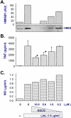
Figure 1. EGCG effectively abrogated endotoxin-induced HMGB1 release in murine macrophage-like RAW 264.7 cells.
RAW 264.7 cells were stimulated with LPS in the absence, or presence of EGCG at indicated concentrations. At 16 hours after LPS stimulation, levels of HMGB1 (Panel A), TNF (Panel B), or nitric oxide (NO, Panel C) in the culture medium were determined by Western blotting, ELISA, and Griess reaction, respectively. Note that at concentrations that completely abrogated LPS-induced HMGB1 release (Panel A), EGCG only partially inhibited LPS-induced TNF secretion (Panel B), but preserved suppress LPS-induced nitric oxide release (Panel C).
Figure 2
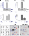
Figure 2. EGCG effectively abrogated endotoxin-induced HMGB1 release in primary macrophage/monocyte cultures.
Primary murine peritoneal macrophages (Panel A, C), or human peripheral blood mononuclear cells (Panel B) were stimulated with LPS in the absence, or presence of EGCG (15 µM). At 16 hours after LPS stimulation, levels of HMGB1 (Panel A, B), nitric oxide (Panel A), TNF (Panel B), or other cytokines (Panel C) in the culture medium were determined by Western blotting analysis (Panel A, B, top), Griess reaction (Panel A, bottom), ELISA (Panel B, bottom), or cytokine array (Panel C), respectively. Note that at concentrations that completely abrogated LPS-induced HMGB1 release, EGCG did not block LPS-induced release of NO (Panel A), G-CSF (Panel C), IL-1α (Panel C), P-selectin (Panel C), or LIX (Panel C). In contrast, EGCG dramatically suppressed LPS-induced release of TNF, sTNF-RII, chemokines (MCP1, MIPs, KC, and RABTES), IL-6, IL-12, and CXCL16 in primary murine macrophages (Panel C). Shown in Panel C was a representative cytokine array of two independent experiments with similar results.
Figure 3
Figure 3. Delayed administration of EGCG still significantly attenuated endotoxin-induced HMGB1 release.
Murine macrophage-like RAW 264.7 cells were stimulated with LPS, and EGCG (10 µM) was added at 0, 2, 6, and 12 hours post LPS stimulation. Levels of HMGB1 levels in the culture medium were determined at 16 hours after LPS stimulation, and expressed (in arbitrary unit, AU) as mean±S.D. of two independent experiments (N = 2). Shown in the lower panel was a representative Western blot. *, P<0.05 versus controls (“+ LPS alone”).
Figure 4
Figure 4. EGCG, but not catechin or ethyl gallate, effectively abrogated endotoxin-induced HMGB1 release.
Macrophage cultures were stimulated with LPS in the absence, or presence of epigallocatechin gallate (EGCG), catechin (C), or ethyl gallate (G) (Panel A), and assayed for HMGB1 release by Western blotting analysis (Panel B) at 16 h post LPS stimulation. Note that epigallocatechin gallate, but not catechin or ethyl gallate, abrogated LPS-induced HMGB1 release.
Figure 5
Figure 5. EGCG significantly protects mice against lethal endotoxemia (Panel A) and lethal sepsis (Panel B).
Balb/C mice were subjected to lethal endotoxemia (LPS, 15 mg/kg, i.p., Panel A), or sepsis (induced by CLP, Panel B). At −0.5, +24, and +48 hours post the onset of endotoxemia, or +24, +48, +72 hours post the onset of sepsis, animals were intraperitoneally administered with saline (0.2 ml/mouse), or EGCG (0.2 ml/mouse, at indicated doses), and animal survival was monitored for up to two weeks. The Kaplan-Meier method was used to compare the differences in mortality rates between groups. *, P<0.05 versus saline.
Figure 6
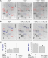
Figure 6. EGCG attenuates sepsis-induced systemic accumulation of IL-6, KC, and HMGB1.
Balb/C mice were subjected to lethal sepsis by CLP, and intraperitoneally administered with control saline (0.2 ml/mouse) or EGCG (4.0 mg/kg) at +24, +48 hours post CLP. At 52 hours post the onset of sepsis, serum levels of cytokines (Panel A, B, C), or HMGB1 (Panel C) were determined by cytokine antibody array (Panel A, B), ELISA (Panel C), or Western blotting analysis (Panel C), respectively. In parallel experiments, serum were pooled from 3 normal mice (-CLP), 3 septic mice approaching moribund state (52 h post CLP), 3 septic mice in non-moribund state (52 h post CLP), and assayed for cytokine profile by antibody array (Panel B). Serum levels of HMGB1 or TNF (Panel C) were expressed as mean±SD (n = 8–10). *, P<0.05 (ANOVA, Tukey test).
Figure 7
Figure 7. EGCG attenuates HMGB1-induced release of proinflammatory mediators.
Murine macrophage-like RAW 264.7 cells (Panel A, Panel C) or primary murine peritoneal macrophages (Panel B) were stimulated with HMGB1 (2.0 µg/ml) in the absence, or presence of EGCG added at indicated concentrations, and time points post HMGB1 stimulation. At 16 hours after HMGB1 stimulation, levels of TNF (Panel A, B, top), IL-6 (Panel C), nitric oxide (Panel A, B, bottom) in the culture medium were determined by ELISA or Griess reaction (Panel A, B), respectively.
Figure 8
Figure 8. EGCG prevents HMGB1 accumulation/clustering on macrophage cell surface.
Macrophage cultures were incubated with biotin-labeled CBP-HMGB1 fusion protein (“+ HMGB1”, 2 μg/ml), in the absence (Panel A), or presence (Panel B, C) of EGCG (10 µM) for various period of time. To visualize exogenous HMGB1, cells were stained with streptavidin-conjugated Alexa 594 (Panel A, “Streptavidin Alexa 594”) or Alexa 488 (Panel B, “Streptavidin Alexa 488” ), or HMGB1-specific rabbit antibodies plus Alexa 488-conjugated goat-anti-rabbit antibodies (Panel A, “Anti-HMGB1 Alexa 488”). Phase contras images indicate macrophage cell morphology; overlay images show co-localization of red and green fluorescence (as yellow). Note anti-HMGB1 antibody-specific immunostaining revealed the presence of both exogenous (on cell surface) and endogenous HMGB1 (in the nucleus) at 4–6 hours post HMGB1 incubation (Panel A, “Anti-HMGB1 Alexa 488”). To determine the relative content of exogenous HMGB1, streptvidin-pulled down fraction or whole cell lysate were immunoblotted with HMGB1-specific antibodies (Panel C). Note EGCG dramatically decreased levels of exogenous HMGB1 (indicated by the 33 kDa band corresponding to CBP-HMGB1 fusion protein) (“rHMGB1”).

References

    1. Riedemann NC, Guo RF, Ward PA. The enigma of sepsis. J Clin Invest. 2003;112:460–467. - PMC - PubMed
    1. Dinarello CA, Thompson RC. Blocking IL-1: interleukin 1 receptor antagonist in vivo and in vitro. Immunol Today. 1991;12:404–410. - PubMed
    1. Yin K, Gribbin E, Wang H. Interferon-gamma inhibition attenuates lethality after cecal ligation and puncture in rats: implication of high mobility group box-1. Shock. 2005;24:396–401. - PubMed
    1. Fink MP, Payen D. The role of nitric oxide in sepsis and ARDS: synopsis of a roundtable conference held in Brussels on 18–20 March 1995. Intensive Care Med. 1996;22:158–165. - PubMed
    1. Vincent JL, Zhang H, Szabo C, Preiser JC. Effects of nitric oxide in septic shock. Am J Respir Crit Care Med. 2000;161:1781–1785. - PubMed

Publication types