. 2008 Jan;19(1):89-96.
doi: 10.1021/bc700161p. Epub 2007 Nov 8.

A versatile bifunctional chelate for radiolabeling humanized anti-CEA antibody with In-111 and Cu-64 at either thiol or amino groups: PET imaging of CEA-positive tumors with whole antibodies

Affiliations

A versatile bifunctional chelate for radiolabeling humanized anti-CEA antibody with In-111 and Cu-64 at either thiol or amino groups: PET imaging of CEA-positive tumors with whole antibodies

Lin Li et al. Bioconjug Chem. 2008 Jan.

Abstract

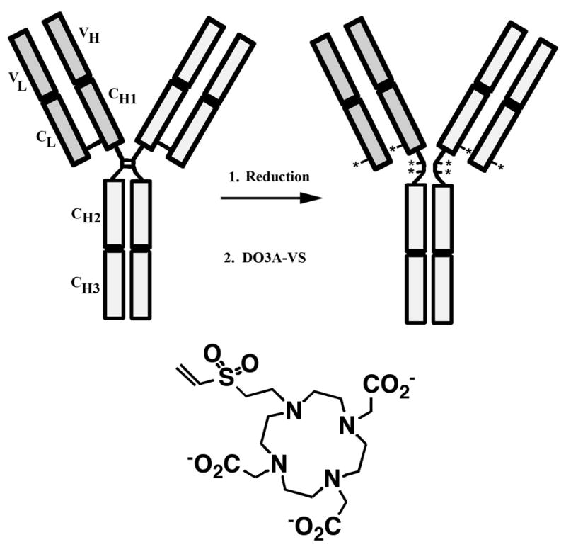
Radiolabeled anti-carcinoembryonic antigen (CEA) antibodies have the potential to give excellent images of a wide variety of human tumors, including tumors of the colon, breast, lung, and medullar thyroid. In order to realize the goals of routine and repetitive clinical imaging with anti-CEA antibodies, it is necessary that the antibodies have a high affinity for CEA, low cross reactivity and uptake in normal tissues, and low immunogenicity. The humanized anti-CEA antibody hT84.66-M5A (M5A) fulfills these criteria with an affinity constant of >10 (10) M (-1), no reactivity with CEA cross-reacting antigens found in normal tissues, and >90% human protein sequence. A further requirement for routine clinical use of radiolabeled antibodies is a versatile method of radiolabeling that allows the use of multiple radionuclides that differ in their radioemissions and half-lives. We describe a versatile bifunctional chelator, DO3A-VS (1,4,7-tris(carboxymethyl)-10-(vinylsulfone)-1,4,7,10-tetraazacyclododecane) that binds a range of radiometals including 111 In for gamma-ray imaging and 64Cu for positron emission tomography (PET), and which can be conjugated with negligible loss of immunoreactivity either to sulfhydryls (SH) in the hinge region of lightly reduced immunoglobulins or surface lysines (NH) of immunoglobulins. Based on our correlative studies comparing the kinetics of radiolabeled anti-CEA antibodies in murine models with those in man, we predict that 64Cu-labeled intact, humanized antibodies can be used to image CEA positive tumors in the clinic.


Figures

Figure 1
Figure 1. Schematic model of an immunoglobulin (IgG1) showing reduction of hinge sulfhydryls
(*). In the second step, the sulfhydryls are reacted with DO3A-VS.
Figure 2
Figure 2. Characterization of DO3A-VS-M5A conjugates by IEF gel electrophoresis
1: IEF standards (see pI values to the left). 2, 5, and 7: unconjugated M5A. 3: M5A after reduction with TCEP. 4: SH-DO3A-VS-M5A. 6: NH-DO3A-VS-M5A. 8: SATA-DO3A-VS-M5A.
Figure 3
Figure 3. MALDI-TOF-MS analysis of SH-DO3A-VS-M5A
A reduced M5A. The peak at 23,893 is the light chain, the peak at 50,672 is the heavy chain and the peaks labeled L2 and HL are light chain and heavy-light chain dimers, respectively. B: SH-DO3A-VS-M5A. The light chain has two peaks, one unmodified at m/z 23,893, and one increased by 462 mass units corresponding to the addition of one DO3A-VS. Based on peak heights the degree of modification for the light chain is 0.67. The heavy chain has increased by 1517 mass units corresponding to the addition of an average 3.3 DO3A-VS. Note: The light chain has one “hinge” cysteine and the heavy chain three hinge cysteine residues in an IgG1 molecule.
Figure 4
Figure 4. SEC analysis of radiolabeled M5A conjugates
A SH-DO3A-VS-M5A radiolabeled with 111In (black) or 64Cu (blue) before and after the addition of NA3 (black dotted) or CEA (blue dotted). B: NH-DO3A-VS-M5A radiolabeled with 111In (black) or 64Cu (blue) before and after the addition of CEA (black or blue dotted). C: SATA-DO3A-VS-M5A radiolabeled with 111In (black) before and after the addition of CEA (black dotted). D: DOTA-NHS-M5A radiolabeled with 111In (black) or 64Cu (blue) before and after the addition of CEA (back or blue dotted). Radioactivity (arbitrary units) shown on the y-axis.
Figure 5
Figure 5. Serum stability analysis of 64Cu-labeled M5A conjugates
64Cu labeled DO3A-VS-M5A was incubated with human serum and analyzed over time by SEC. The major peak at 42 min corresponds to the elution time for IgG. A: SH-DO3A-VS-M5A. B: NH-DO3A-VS-M5A. The sample was also injected into nude mice, blood removed over time, and the serum fraction analyzed by SEC. C: SH-DO3A-VS-M5A. D: NH-DO3A-VS-M5A.
Figure 6
Figure 6. Biodistributions of 111In- and 64Cu-SH- and NH-DO3A-VS-M5A in nude mice bearing LS174T xenografts
The average %ID/g (± sem.) of five animals per time point is shown. A: 111In-labeled SH-DO3A-VS-M5A. B: 64Cu-labeled SH-DO3A-VS-M5A. C: 111In-labeled NH-DO3A-VS-M5A. D: 64Cu-labeled NH-DO3A-VS-M5A.
Figure 7
Figure 7. Biodistributions of 111In- SATA-DO3A-VS-M5A and DOTA-NHS-M5A in nude mice bearing LS174T xenografts
The average %ID/g (± sem) of five animals per time point is shown. A: 111In-labeled SATA-DO3A-VS-M5A. B: 111In-labeled DOTA-NHS-M5A.
Figure 8
Figure 8. Dynamic biodistribution of 64Cu-labeled hT84.66-M5A MAbs in athymic mice peripherally xenografted with CEA-positive LS-174T colon tumors
(A) SH-DO3A-VS-M5A; (B) NH-DO3A-VS-M5A. Images are PET anterior-view maximum intensity projections (MIPs) normalized to reflect radiodecay-corrected relative image intensity per unit injected activity. Labels shown in red, yellow and green show directly-measured %ID/g in blood (heart), liver and tumor, respectively. Tumor weights at time of sacrifice were 126 and 155 mg in (A) and (B) respectively.
Figure 9
Figure 9. Time-activity curves derived from serial microPET studies
Data are average %ID/g (± sem.) for three (first 3 time points for SH-DO3A-VS-M5A) or two (NH-DO3A-VS-M5A and final time point for SH-DO3A-VS-M5A) mice.

References

    1. Williams LE, Wu AM, Yazaki PJ, Liu A, Raubitschek AA, Shively JE, Wong JY. Numerical selection of optimal tumor imaging agents with application to engineered antibodies. Cancer Biother Radiopharm. 2001;16:25–35. - PubMed
    1. Kenanova V, Wu AM. Tailoring antibodies for radionuclide delivery. Expert Opin Drug Deliv. 2006;3:53–70. - PubMed
    1. Yazaki PJ, Wu AM, Tsai SW, Williams LE, Ikler DN, Wong JY, Shively JE, Raubitschek AA. Tumor targeting of radiometal labeled anti-CEA recombinant T84.66 diabody and T84.66 minibody: comparison to radioiodinated fragments. Bioconjug Chem. 2001;12:220–8. - PubMed
    1. Li L, Yazaki PJ, Anderson AL, Crow D, Colcher D, Wu AM, Williams LE, Wong JY, Raubitschek A, Shively JE. Improved biodistribution and radioimmunoimaging with poly(ethylene glycol)-DOTA-conjugated anti-CEA diabody. Bioconjug Chem. 2006;17:68–76. - PubMed
    1. Milenic DE, Garmestani K, Chappell LL, Dadachova E, Yordanov A, Ma D, Schlom J, Brechbiel MW. In vivo comparison of macrocyclic and acyclic ligands for radiolabeling of monoclonal antibodies with 177Lu for radioimmunotherapeutic applications. Nucl Med Biol. 2002;29:431–42. - PubMed

Publication types