. 2007 Nov 8;56(3):503-16.
doi: 10.1016/j.neuron.2007.10.024.

Two Gr genes underlie sugar reception in Drosophila

Affiliations

Two Gr genes underlie sugar reception in Drosophila

Anupama Dahanukar et al. Neuron. .

Abstract

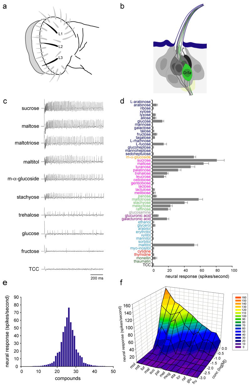
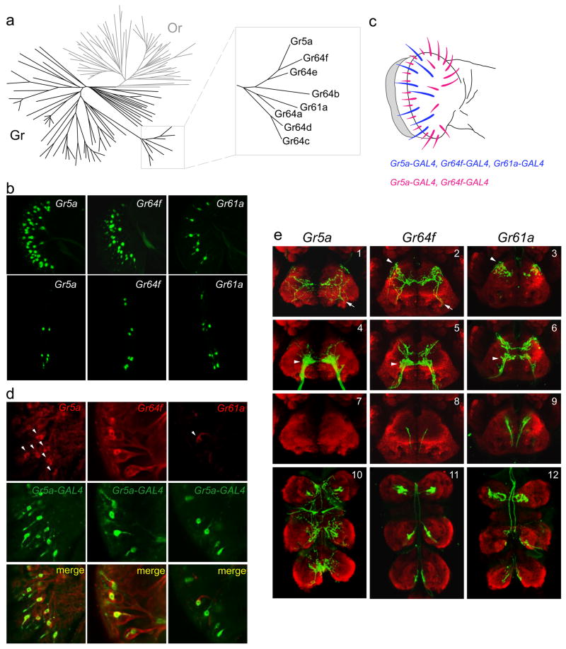
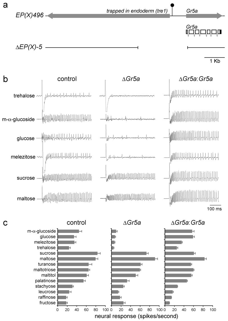
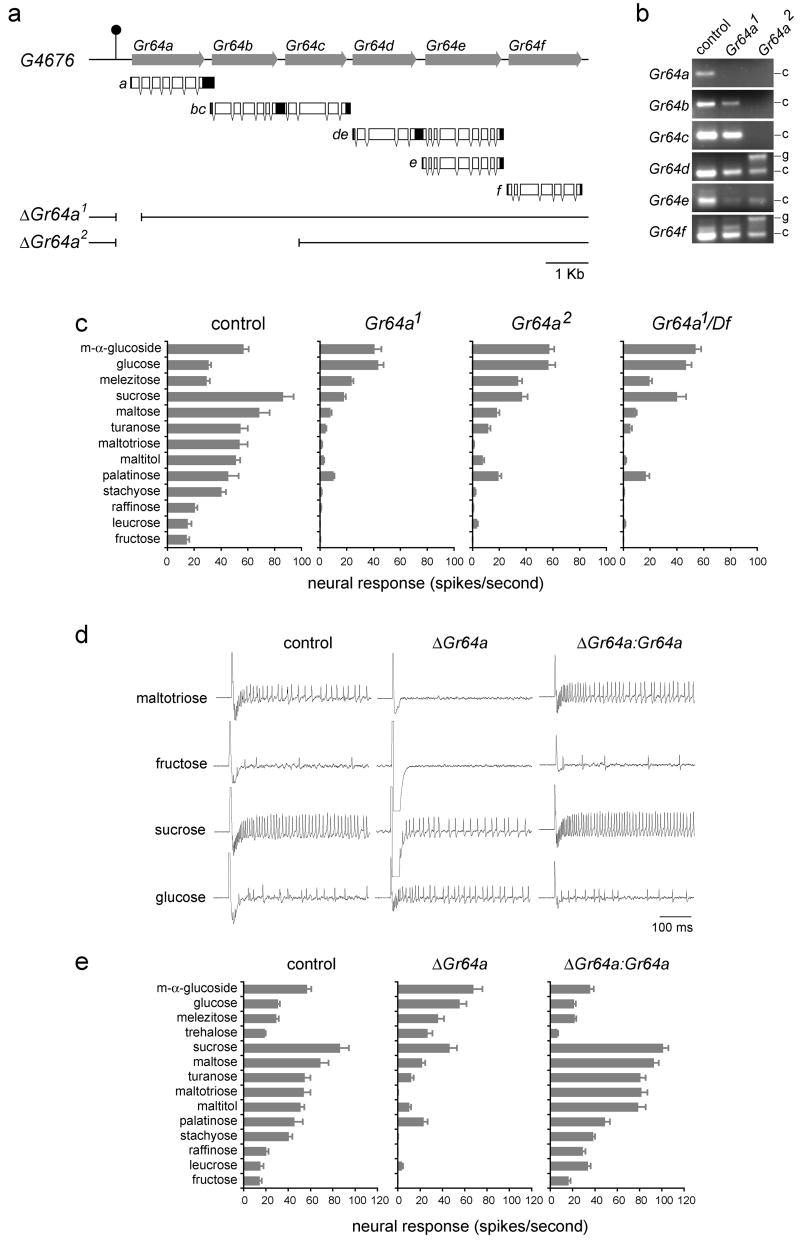
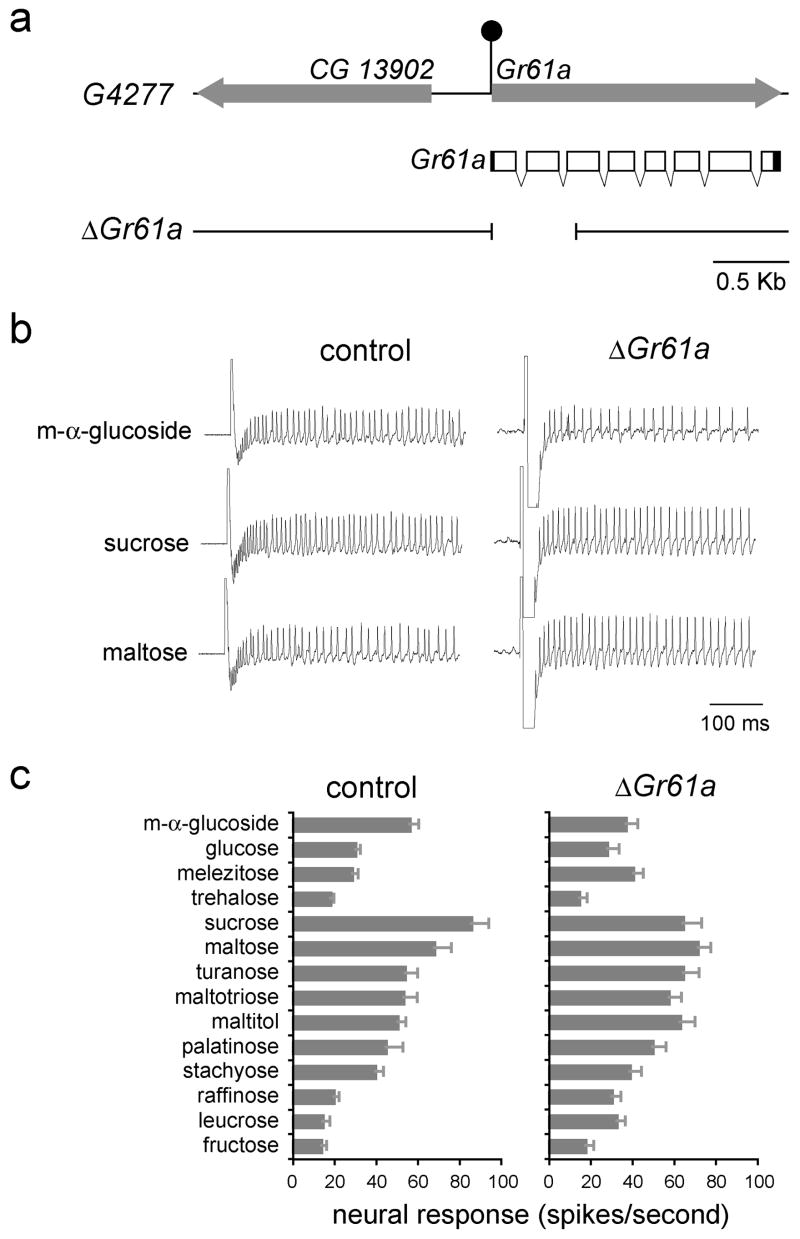
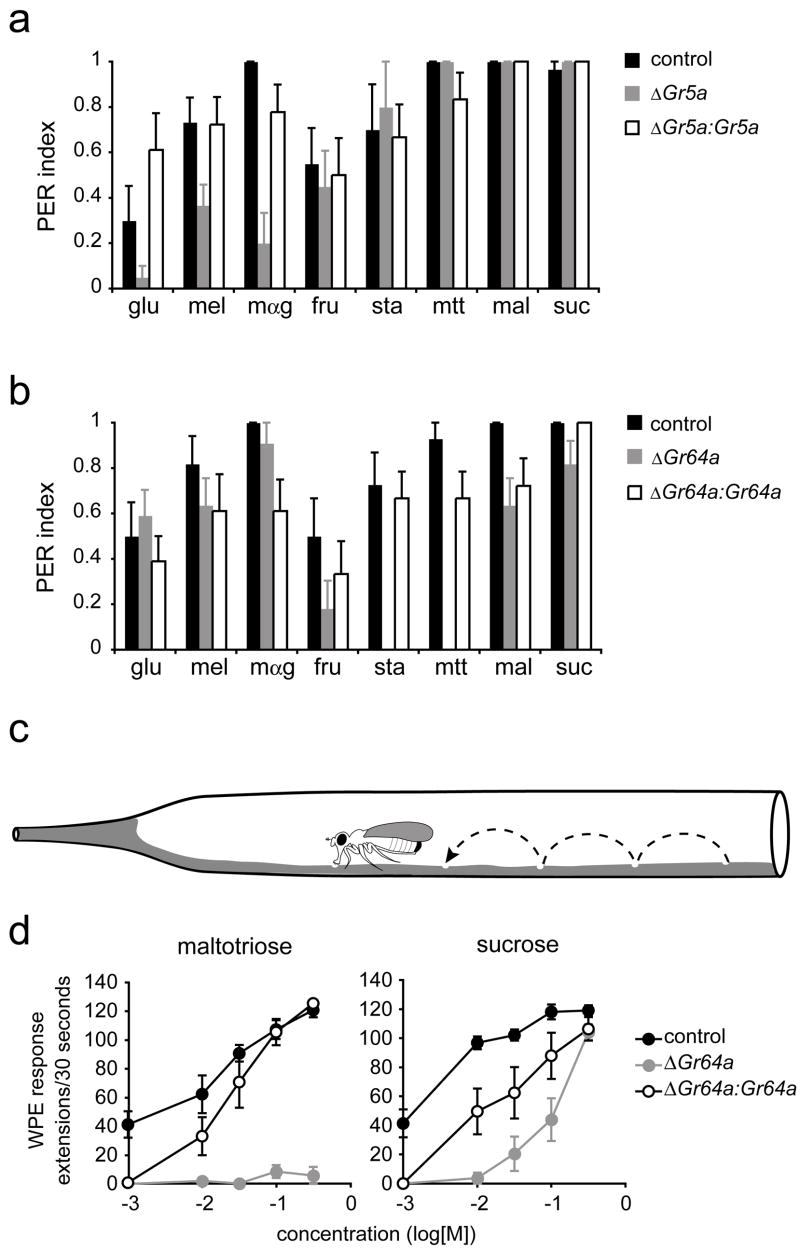
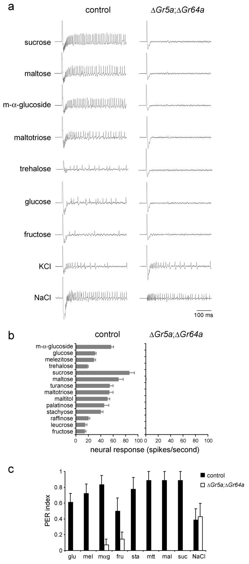
We have analyzed the molecular basis of sugar reception in Drosophila. We define the response spectrum, concentration dependence, and temporal dynamics of sugar-sensing neurons. Using in situ hybridization and reporter gene expression, we identify members of the Gr5a-related taste receptor subfamily that are coexpressed in sugar neurons. Neurons expressing reporters of different Gr5a-related genes send overlapping but distinct projections to the brain and thoracic ganglia. Genetic analysis of receptor genes shows that Gr5a is required for response to one subset of sugars and Gr64a for response to a complementary subset. A Gr5a;Gr64a double mutant shows no physiological or behavioral responses to any tested sugar. The simplest interpretation of our results is that Gr5a and Gr64a are each capable of functioning independently of each other within individual sugar neurons and that they are the primary receptors used in the labellum to detect sugars.


Figures

Figure 1
Figure 1. Responses of sugar neurons
(a) Labellar sensilla. The three L-type sensilla are in black. (b) An L-type sensillum, showing the sugar neuron (green), other gustatory neurons (dark gray), the mechanosensory neuron (black) and supporting cells (light gray). (c) Sample traces of recordings from L-type sensilla. A control trace is shown using the diluent, tri-choline citrate (TCC), alone. (d) Responses to a panel of 50 compounds. Sugars were D-isomers except as indicated. Chemicals are color coded: monosaccharides (dark blue), a glucoside (orange), disaccharides (pink), oligosaccharides (light green), glucosamine (gray), sugar acids (violet), alcohols (light blue), nucleotides (red), proteins (dark green), diluent control (black). All compounds were tested at a concentration of 100 mM except for ethanol (25% v/v), monellin (0.1% w/v), thaumatin (0.1% w/v) and TCC (30 mM). For all stimuli, 10≤n≤15. Error bars indicate SEM. (e) Tuning curve for L-type sensilla. The 50 stimuli are arranged along the X-axis according to the strengths of the responses that they elicit. Those that elicit the strongest responses are placed near the center, and those that elicit the weakest responses are placed near the edges. (f) Concentration-dependent responses to a panel of 13 sugars. n=6. Maltitol (mol), maltotriose (mtt), turanose (tur), maltose (mal), sucrose (suc), palatinose (pal), melizitose (mel), m-α-glucoside (mαg), stachyose (sta), leucrose (lcr), raffinose (raf), glucose (glu) fructose (fru).
Figure 2
Figure 2. Expression of Gr5a-related receptors
(a) Phylogenetic tree depicting the chemoreceptor superfamily in Drosophila, indicating odor receptors (Or) in gray and gustatory receptors (Gr) in black. Inset shows the Gr5a sub-family. Adapted from van der Goes van Naters and Carlson (2006). (b) GFP reporter expression in the labellum (top) or the three distal-most segments of forelegs (bottom), driven by Gr5a-GAL4 (left), Gr64f-GAL4 (center) or Gr61a-GAL4 (right). (c) A receptor-to-sensillum map of Gr5a, Gr64f and Gr61a based on the data represented in (b). (d) In situ hybridizations with probes against the indicated mRNAs (red) to labella of Gr5a-GAL4;UAS-GFP flies that were simultaneously stained for GFP (green). (e) Axonal projections of neurons labeled by the indicated drivers (green). The neuropil is stained with nc82 (red). Shown here are optical sections of anterior views of the SOG (1–9, dorsal is up) and the thoracic ganglion (10–12, anterior is up).
Figure 3
Figure 3. The trehalose receptor, Gr5a, mediates responses to several sugars
(a) The Gr5a genomic region. The filled arrows indicate the trapped in endoderm (Tre1) and Gr5a transcription units. The filled circle depicts the P element insertion in EP(X)496. Shown below is the transcript structure for Gr5a, indicating both protein-coding (hollow) and untranslated (filled) regions. Sequences deleted in ΔEP(X)-5 are indicated at the bottom. (b) Sample traces from the indicated genotypes. (c) Sugar response profiles of L-type sensilla from EP(X)496 (control), ΔEP(X)-5Gr5a) or from ΔEP(X)-5;Gr5a-GAL4;UAS-Gr5aGr5a:Gr5a) flies. Sugars were tested at 100 mM, except for glucose and fructose, which were tested at 300 mM. For all graphs, n=10–11. Error bars indicate SEM.
Figure 4
Figure 4. Gr64a mediates responses to several sugars
(a) The Gr64a-Gr64f genomic region. The filled arrows indicate the Gr64a-Gr64f genes; transcript structures are shown below indicating protein-coding (hollow) and untranslated (filled) regions. The filled circle depicts the P element insertion site in G4676. Sequences deleted in Gr64a1 and Gr64a2 are indicated below. (b) RT-PCR expression analysis of Gr64a-Gr64f in precise excision (control), Gr64a1/Gr64a1, and Gr64a2/Gr64a2 flies as indicated. The positions of the cDNA (c) and genomic (g) products are indicated on the right. All products agree with the predicted sizes. (c) Sugar response profiles of L-type sensilla from precise excision (control), Gr64a1/Gr64a1, Gr64a2/Gr64a2, and Gr64a1/Df(3L)GN34 male flies. (d) Sample traces from the indicated genotypes. (e) Sugar response profiles of L-type sensilla from precise excision (control), Gr64a1/Gr64a2Gr64a), and Gr5a-GAL4,UAS-Gr64a/UAS-Gr64a;Gr64a1/Gr64a2Gr64a:Gr64a). Sugars were tested at 100 mM, except for glucose and fructose, which were tested at 300 mM. n=9–12. Error bars indicate SEM.
Figure 5
Figure 5. Most sugar responses are not reduced by loss of Gr61a
(a) The Gr61a genomic region. The filled arrows indicate transcription units. Shown below is the transcript structure for Gr61a, indicating both protein-coding (hollow) and untranslated (filled) regions. The filled circle depicts the P element insertion in G4277. Sequences deleted in ΔGr61a are indicated below. (b) Representative traces from control flies and Gr61a1/Gr61a1Gr61a) flies. (c) Sugar response profiles of L-type sensilla from control and Gr61a1/Gr61a1Gr61a) flies. Sugars were tested at 100 mM, except for glucose and fructose, which were tested at 300 mM. n=9–10. Error bars indicate SEM.
Figure 6
Figure 6. Gr5a and Gr64a mediate behavioral responses to sugars
(a,b) Proboscis extension responses. (a) EP(X)496 (control), ΔEP(X)-5Gr5a) and ΔEP(X)- 5;Gr5a-GAL4;UAS-Gr5aGr5a:Gr5a); n=8–15 for all sugars except for stachyose, n=5–9. (b) Precise excision (control), Gr64a1/Gr64a1Gr64a) and Gr5a-GAL4/UAS-Gr64a;Gr64a1/Gr64a1Gr64a:Gr64a). n=9–14. Error bars indicate SEM. All sugars were tested at 100 mM. glucose (glu), melezitose (mel), m-α-glucoside (mαg), fructose (fru), stachyose (sta), maltotriose (mtt), maltose (mal), sucrose (suc). (c) The walking proboscis extension (WPE) assay. Not drawn to scale. (d) Proboscis extensions in the walking assay. Genotypes are: precise excision (control), Gr64a1/Gr64a2Gr64a) and Gr5a-GAL4/UAS-Gr64a;Gr64a1/Gr64a2Gr64a:Gr64a). n=9–10. Error bars indicate SEM.
Figure 7
Figure 7. Labellar sugar responses depend on either Gr5a or Gr64a
(a)Sample traces of physiological recordings from precise excision (control), or ΔEP(X)-5;Gr64a2/Gr64a2Gr5a; ΔGr64a) flies as indicated. (b) Response profiles. All sugars were tested at 100 mM, except for glucose and fructose, which were tested at 300 mM. KCl was tested at 1 mM, and NaCl at 400 mM. n=9–12 for control, n=6 for mutants. Error bars indicate SEM. (c) Proboscis extension responses of control flies (precise excision), or ΔGr5a; ΔGr64a (ΔEP(X)-5;Gr64a2/Gr64a2) flies as indicated. None of the responses of the mutant exceeded a control response to pure water (p<0.05). All sugars were tested at 100 mM. glucose (glu), melezitose (mel), m-α-glucoside (mαg), fructose (fru), stachyose (sta), maltotriose (mtt), maltose (mal), sucrose (suc). NaCl was tested at 5 mM. n=7–9. Error bars indicate SEM.

References

    1. Benton R, Sachse S, Michnick SW, Vosshall LB. Atypical membrane topology and heteromeric function of Drosophila odorant receptors in vivo. PLoS Biol. 2006;4:e20. - PMC - PubMed
    1. Brand AH, Perrimon N. Targeted gene expression as a means of altering cell fates and generating dominant phenotypes. Development. 1993;118:401–415. - PubMed
    1. Cheng E, Chang C-M, Sun HY. An improved proboscis extension assay. Drosophila. Information Newsletter. 1992;6
    1. Chyb S, Dahanukar A, Wickens A, Carlson JR. Drosophila Gr5a encodes a taste receptor tuned to trehalose. Proc Natl Acad Sci U S A. 2003;100:14526–14530. - PMC - PubMed
    1. Clyne PJ, Warr CG, Carlson JR. Candidate taste receptors in Drosophila. Science. 2000;287:1830–1834. - PubMed

Publication types

MeSH terms