

Assessment of functional development in normal infant brain using arterial spin labeled perfusion MRI
- PMID: 17988892
- PMCID: PMC2268607
- DOI: 10.1016/j.neuroimage.2007.09.045
Assessment of functional development in normal infant brain using arterial spin labeled perfusion MRI
Abstract
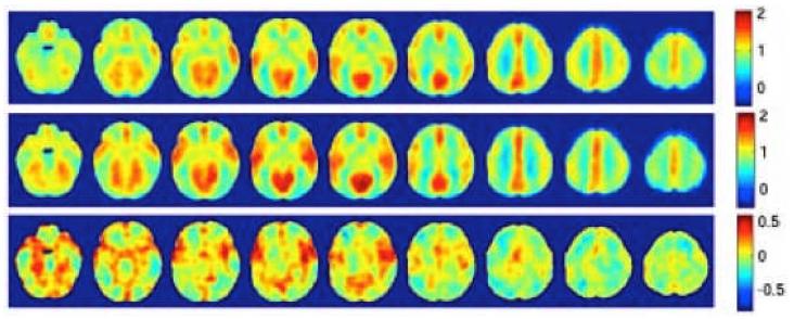
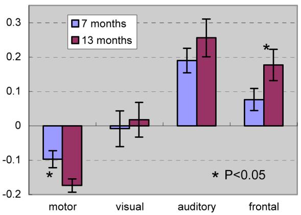
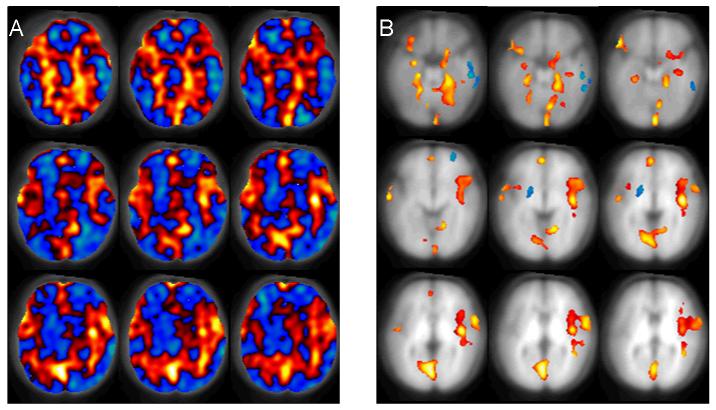
Arterial spin labeled (ASL) perfusion MRI provides a noninvasive approach for longitudinal imaging of regional brain function in infants. In the present study, continuous ASL (CASL) perfusion MRI was carried out in normally developing 7- and 13-month-old infants while asleep without sedation. The 13-month infant group showed an increase (P<0.05) of relative CBF in frontal regions as compared to the 7-month group using a region of interest based analysis. Using a machine-learning algorithm to automatically classify the relative CBF maps of the two infant groups, a significant (P<0.05, permutation testing) regional CBF increase was found in the hippocampi, anterior cingulate, amygdalae, occipital lobes, and auditory cortex in the 13-month-old infants. These results are consistent with current understanding of infant brain development and demonstrate the feasibility of using perfusion MRI to noninvasively monitor developing brain function.
Figures
Similar articles
-
Heterogeneous increases of regional cerebral blood flow during preterm brain development: Preliminary assessment with pseudo-continuous arterial spin labeled perfusion MRI.Neuroimage. 2017 Feb 15;147:233-242. doi: 10.1016/j.neuroimage.2016.12.034. Epub 2016 Dec 14. Neuroimage. 2017. PMID: 27988320 Free PMC article.
-
Multi-phase 3D arterial spin labeling brain MRI in assessing cerebral blood perfusion and arterial transit times in children at 3T.Clin Imaging. 2019 Jan-Feb;53:210-220. doi: 10.1016/j.clinimag.2018.11.001. Epub 2018 Nov 6. Clin Imaging. 2019. PMID: 30439588
-
Regional and depth-dependence of cortical blood-flow assessed with high-resolution Arterial Spin Labeling (ASL).J Cereb Blood Flow Metab. 2021 Aug;41(8):1899-1911. doi: 10.1177/0271678X20982382. Epub 2021 Jan 14. J Cereb Blood Flow Metab. 2021. PMID: 33444098 Free PMC article.
-
Arterial spin-labeled perfusion MRI in basic and clinical neuroscience.Curr Opin Neurol. 2009 Aug;22(4):348-55. doi: 10.1097/WCO.0b013e32832d9505. Curr Opin Neurol. 2009. PMID: 19491678 Review.
-
Mapping of cerebral perfusion territories using territorial arterial spin labeling: techniques and clinical application.NMR Biomed. 2013 Aug;26(8):901-12. doi: 10.1002/nbm.2836. Epub 2012 Jul 15. NMR Biomed. 2013. PMID: 22807022 Review.
Cited by
-
Multidelay Arterial Spin-Labeling MRI in Neonates and Infants: Cerebral Perfusion Changes during Brain Maturation.AJNR Am J Neuroradiol. 2018 Oct;39(10):1912-1918. doi: 10.3174/ajnr.A5774. Epub 2018 Sep 13. AJNR Am J Neuroradiol. 2018. PMID: 30213808 Free PMC article.
-
Multi-task prediction of infant cognitive scores from longitudinal incomplete neuroimaging data.Neuroimage. 2019 Jan 15;185:783-792. doi: 10.1016/j.neuroimage.2018.04.052. Epub 2018 Apr 27. Neuroimage. 2019. PMID: 29709627 Free PMC article.
-
Spatiotemporal cerebral blood flow dynamics underlies emergence of the limbic-sensorimotor-association cortical gradient in human infancy.Res Sq [Preprint]. 2024 Aug 8:rs.3.rs-4761517. doi: 10.21203/rs.3.rs-4761517/v1. Res Sq. 2024. Update in: Nat Commun. 2024 Oct 17;15(1):8944. doi: 10.1038/s41467-024-53354-7. PMID: 39149463 Free PMC article. Updated. Preprint.
-
Heterogeneous increases of regional cerebral blood flow during preterm brain development: Preliminary assessment with pseudo-continuous arterial spin labeled perfusion MRI.Neuroimage. 2017 Feb 15;147:233-242. doi: 10.1016/j.neuroimage.2016.12.034. Epub 2016 Dec 14. Neuroimage. 2017. PMID: 27988320 Free PMC article.
-
Rest Functional Brain Maturation during the First Year of Life.Cereb Cortex. 2021 Feb 5;31(3):1776-1785. doi: 10.1093/cercor/bhaa325. Cereb Cortex. 2021. PMID: 33230520 Free PMC article.
References
-
- Alsop DC, Detre JA. Multisection Cerebral Blood Flow MR Imaging with Continuous Arterial Spin Labeling. Radiology. 1998;208:410–416. - PubMed
-
- Alsop DC, Detre JA, Grossman M. Assessment of cerebral blood flow in Alzheimer's disease by spin-labeled magnetic resonance imaging. Annals of neurology. 2000;47:93–100. - PubMed
-
- Biagi L, Abbruzzese A, Bianchi MC, Alsop DC, Guerra AD, Tosetti M. Age dependence of cerebral perfusion assessed by magnetic resonance continuous arterial spin labeling. J Magn Reson Imaging. 2007;25(4):696–702. - PubMed
-
- Biswal BB, Ulmer JL. Blind source separation of multiple signal sources of fMRI data sets using independent component analysis. Journal of Computer Assisted Tomography. 1999;23:265–271. - PubMed
-
- Burges CJC. A Tutorial on Support Vector Machines for Pattern Recognition. Data Mining and Knowledge Discovery. 1998;2:121–167.
Publication types
MeSH terms
Substances
Grants and funding
LinkOut - more resources
Full Text Sources
