Review
. 2008 Mar 27;363(1494):1167-77; discussion 1177.
doi: 10.1098/rstb.2007.2212.

Focusing the view on nature's water-splitting catalyst

Affiliations
Review

Focusing the view on nature's water-splitting catalyst

Samir Zein et al. Philos Trans R Soc Lond B Biol Sci. .

Abstract

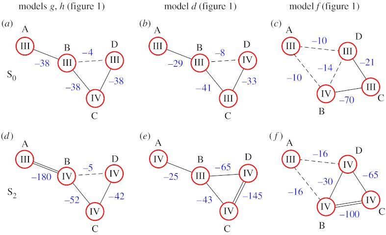
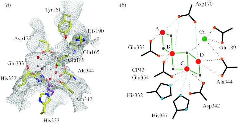
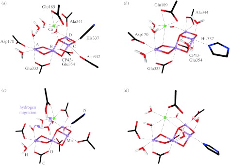
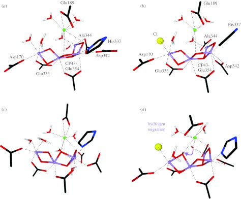
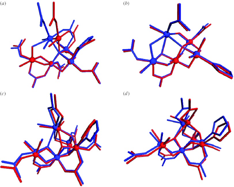
Nature invented a catalyst about 3Gyr ago, which splits water with high efficiency into molecular oxygen and hydrogen equivalents (protons and electrons). This reaction is energetically driven by sunlight and the active centre contains relatively cheap and abundant metals: manganese and calcium. This biological system therefore forms the paradigm for all man-made attempts for direct solar fuel production, and several studies are underway to determine the electronic and geometric structures of this catalyst. In this report we briefly summarize the problems and the current status of these efforts and propose a density functional theory-based strategy for obtaining a reliable high-resolution structure of this unique catalyst that includes both the inorganic core and the first ligand sphere.


Figures

Figure 1
Figure 1
(ah) Proposed models for the inorganic core of the WOC. Red spheres symbolize manganese ions, blue spheres oxygen bridges and green spheres calcium ions. Dashed lines indicate that the bridging motif is unknown. Models redrawn from DeRose et al. (1994), Ferreira et al. (2004) and Yano et al. (2006). Models g and h correspond to models II and III in Yano et al. (2006), respectively.
Figure 2
Figure 2
Proposed spin-coupling schemes for the (a, b, c) S0 and (d, e, f) S2 states of the WOC, which are consistent with Q-band 55Mn-ENDOR data (Kulik et al. 2005). Red circles indicate the Mn ions, which are labelled A, B, C and D in the same way as in figure 1. The roman numbers in the circles give the formal oxidation states of the Mn ions. Calcium is not included in the spin-coupling schemes since coupling between Mn ions via Ca is assumed to be negligible. The blue numbers give the J-couplings in cm−1 according to H=Jik(SiSk). The type of line represents the coupling strength: double line, strong antiferromagnetic coupling; single solid line, medium strength antiferromagnetic coupling; and dashed lines, weak ferro- or antiferromagnetic coupling.
Figure 3
Figure 3
Ligand environment of model g of figure 1 as determined by placing this model into the 3.0 Å crystal structure (Loll et al. 2005; Yano et al. 2006). (a) Electron density with model g. (b) Schematic of the WOC. Blue dashed lines indicate that the displayed amino acids are too far away (more than 3 Å) to be normally considered as ligands. Dashed black lines indicate metal–ligand distances of less than 3 Å. Mn ions are indicated by red spheres, bridging oxygen by grey spheres and Ca by a green sphere. Ligand oxygens are marked orange and ligand nitrogens blue. For further details, see Yano et al. (2006).
Figure 4
Figure 4
Comparison of the (a,b) initial structures of models II and IIa, with those obtained (c,d) after constrained optimization. All indicated amino acids are from the reaction centre D1 protein of PSII, with the exception of Glu354, which is a side chain of the inner antenna CP43 protein of PSII. Alanine 344 is the C-terminus of the D1 protein and ligates via its terminal carboxy group. Manganese ions are indicated in purple, oxygen in red, hydrogen in white, calcium in green and carbon in black.
Figure 5
Figure 5
Comparison of the (a,b) initial structures of models III and III–Cl with those obtained (c,d) after constrained optimization. All indicated amino acids are from the reaction centre D1 protein of PSII, with the exception of Glu354, which is a side chain of the inner antenna CP43 protein of PSII. Alanine 344 is the C-terminus of the D1 protein and ligates via its terminal carboxy group. Manganese ions are indicated in purple, oxygen in red, hydrogen in white, calcium in green, chloride in yellow, carbon in black and nitrogen in blue.
Figure 6
Figure 6
Molecular structures of (a) II, (b) IIa, (c) III and (d) III–Cl models after constrained optimization (red) and after full optimization (blue). Manganese and calcium ions are represented by spheres. All hydrogen atoms are removed for clarity.

References

    1. Becke A.D. Density-functional exchange-energy approximation with correct asymptotic-behavior. Phys. Rev. A. 1988;38:3098–3100. doi:10.1103/PhysRevA.38.3098 - DOI - PubMed
    1. Becke A.D. A new mixing of Hartree–Fock and local density-functional theories. J. Chem. Phys. 1993a;98:1372–1377. doi:10.1063/1.464304 - DOI
    1. Becke A.D. Density-functional thermochemistry. 3. The role of exact exchange. J. Chem. Phys. 1993b;98:5648–5652. doi:10.1063/1.464913 - DOI
    1. Blomberg M.R.A, Siegbahn P.E.M, Styring S, Babcock G.T, Åkermark B, Korall P. A quantum chemical study of hydrogen abstraction from manganese coordinated water by a tyrosyl radical: a model for water oxidation in photosystem II. J. Am. Chem. Soc. 1997;119:8285–8292. doi:10.1021/ja9642323 - DOI
    1. Britt R.D, Peloquin J.M, Campbell K.A. Pulsed and parallel-polarization EPR characterization of the photosystem II oxygen evolving complex. Annu. Rev. Biophys. Biomol. Struct. 2000;29:463–495. doi:10.1146/annurev.biophys.29.1.463 - DOI - PubMed

Additional references

    1. Boussac A, Kuhl H, Ghibaudi E, Rögner M, Rutherford A.W. Detection of an electron paramagnetic resonance signal in the S0 state of the manganese complex of photosystem II from Synechococcus elongatus. Biochemistry. 1999;38:11 942–11 948. doi:10.1021/bi990845r - DOI - PubMed
    1. Charlot M.F, Boussac A, Blondin G. Towards a spin coupling model for the Mn4 cluster in photosystem II. Biochim. Biophys. Acta. 2005;1708:120–132. doi:10.1016/j.bbabio.2005.01.006 - DOI - PubMed
    1. Messinger J, Robblee J.H, Yu W.O, Sauer K, Yachandra V.K, Klein M.P. The S0 state of the oxygen evolving complex in photosystem II is paramagnetic: detection of an EPR multiline signal. J. Am. Chem. Soc. 1997;119:11 349–11 350. doi:10.1021/ja972696a - DOI - PMC - PubMed

Publication types