

The Stanford Tissue Microarray Database
- PMID: 17989087
- PMCID: PMC2238948
- DOI: 10.1093/nar/gkm861
The Stanford Tissue Microarray Database
Abstract
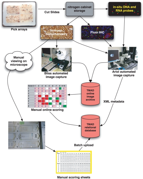
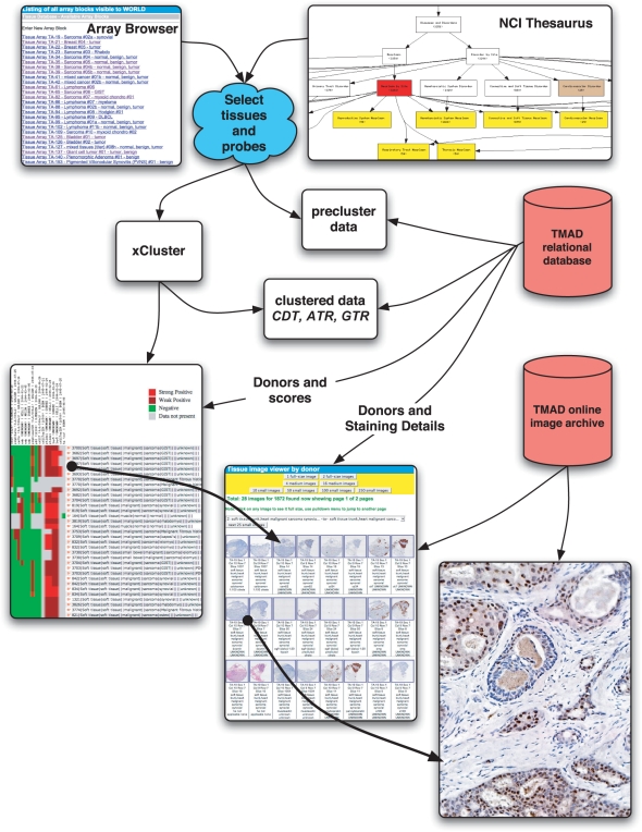
The Stanford Tissue Microarray Database (TMAD; http://tma.stanford.edu) is a public resource for disseminating annotated tissue images and associated expression data. Stanford University pathologists, researchers and their collaborators worldwide use TMAD for designing, viewing, scoring and analyzing their tissue microarrays. The use of tissue microarrays allows hundreds of human tissue cores to be simultaneously probed by antibodies to detect protein abundance (Immunohistochemistry; IHC), or by labeled nucleic acids (in situ hybridization; ISH) to detect transcript abundance. TMAD archives multi-wavelength fluorescence and bright-field images of tissue microarrays for scoring and analysis. As of July 2007, TMAD contained 205 161 images archiving 349 distinct probes on 1488 tissue microarray slides. Of these, 31 306 images for 68 probes on 125 slides have been released to the public. To date, 12 publications have been based on these raw public data. TMAD incorporates the NCI Thesaurus ontology for searching tissues in the cancer domain. Image processing researchers can extract images and scores for training and testing classification algorithms. The production server uses the Apache HTTP Server, Oracle Database and Perl application code. Source code is available to interested researchers under a no-cost license.
Figures

Similar articles
-
The Stanford Microarray Database: implementation of new analysis tools and open source release of software.Nucleic Acids Res. 2007 Jan;35(Database issue):D766-70. doi: 10.1093/nar/gkl1019. Epub 2006 Dec 20. Nucleic Acids Res. 2007. PMID: 17182626 Free PMC article.
-
Annotation and query of tissue microarray data using the NCI Thesaurus.BMC Bioinformatics. 2007 Aug 8;8:296. doi: 10.1186/1471-2105-8-296. BMC Bioinformatics. 2007. PMID: 17686183 Free PMC article.
-
The Stanford Microarray Database accommodates additional microarray platforms and data formats.Nucleic Acids Res. 2005 Jan 1;33(Database issue):D580-2. doi: 10.1093/nar/gki006. Nucleic Acids Res. 2005. PMID: 15608265 Free PMC article.
-
Post-genomic applications of tissue microarrays: basic research, prognostic oncology, clinical genomics and drug discovery.Histol Histopathol. 2004 Jan;19(1):325-35. doi: 10.14670/HH-19.325. Histol Histopathol. 2004. PMID: 14702201 Review.
-
Virtual microscopy as an enabler of automated/quantitative assessment of protein expression in TMAs.Histochem Cell Biol. 2008 Sep;130(3):447-63. doi: 10.1007/s00418-008-0480-1. Epub 2008 Aug 5. Histochem Cell Biol. 2008. PMID: 18679705 Review.
Cited by
-
TIGIT is Frequently Expressed in the Tumor Microenvironment of Select Lymphomas: Implications for Targeted Therapy.Am J Surg Pathol. 2024 Mar 1;48(3):337-352. doi: 10.1097/PAS.0000000000002168. Epub 2023 Dec 22. Am J Surg Pathol. 2024. PMID: 38148663 Free PMC article.
-
KLF1/EKLF expression in acute leukemia is correlated with chromosomal abnormalities.Blood Cells Mol Dis. 2020 Jul;83:102434. doi: 10.1016/j.bcmd.2020.102434. Epub 2020 Apr 13. Blood Cells Mol Dis. 2020. PMID: 32311573 Free PMC article.
-
Multi-modal analysis reveals tumor and immune features distinguishing EBV-positive and EBV-negative post-transplant lymphoproliferative disorders.Cell Rep Med. 2024 Dec 17;5(12):101851. doi: 10.1016/j.xcrm.2024.101851. Epub 2024 Dec 9. Cell Rep Med. 2024. PMID: 39657667 Free PMC article.
-
Systematic planning of genome-scale experiments in poorly studied species.PLoS Comput Biol. 2010 Mar 5;6(3):e1000698. doi: 10.1371/journal.pcbi.1000698. PLoS Comput Biol. 2010. PMID: 20221257 Free PMC article.
-
Harnessing multimodal data integration to advance precision oncology.Nat Rev Cancer. 2022 Feb;22(2):114-126. doi: 10.1038/s41568-021-00408-3. Epub 2021 Oct 18. Nat Rev Cancer. 2022. PMID: 34663944 Free PMC article. Review.
References
-
- Wan W, Fortuna M, Furmanski P. A rapid and efficient method for testing immunohistochemical reactivity of monoclonal antibodies against multiple tissue samples simultaneously. J. Immunol. Methods. 1987;103:121–129. - PubMed
-
- Kononen J, Bubendorf L, Kallioniemi A, Bärlund M, Schraml P, Leighton S, Torhorst J, Mihatsch M, Sauter G, et al. Tissue microarrays for high-throughput molecular profiling of tumor specimens. Nat. Med. 1998;4:844–847. - PubMed
-
- Lee H, Park Y, Sim J, Park R, Kim W, Kim J. The tissue microarray object model: a data model for storage, analysis, and exchange of tissue microarray experimental data. Arch. Pathol. Lab. Med. 2006;130:1004–1013. - PubMed
Publication types
MeSH terms
Substances
Grants and funding
LinkOut - more resources
Full Text Sources
Other Literature Sources
Research Materials
