. 2007 Nov 13;104(46):18321-6.
doi: 10.1073/pnas.0709096104. Epub 2007 Nov 7.

Activating mutation in a mucolipin transient receptor potential channel leads to melanocyte loss in varitint-waddler mice

Affiliations

Activating mutation in a mucolipin transient receptor potential channel leads to melanocyte loss in varitint-waddler mice

Haoxing Xu et al. Proc Natl Acad Sci U S A. .

Abstract

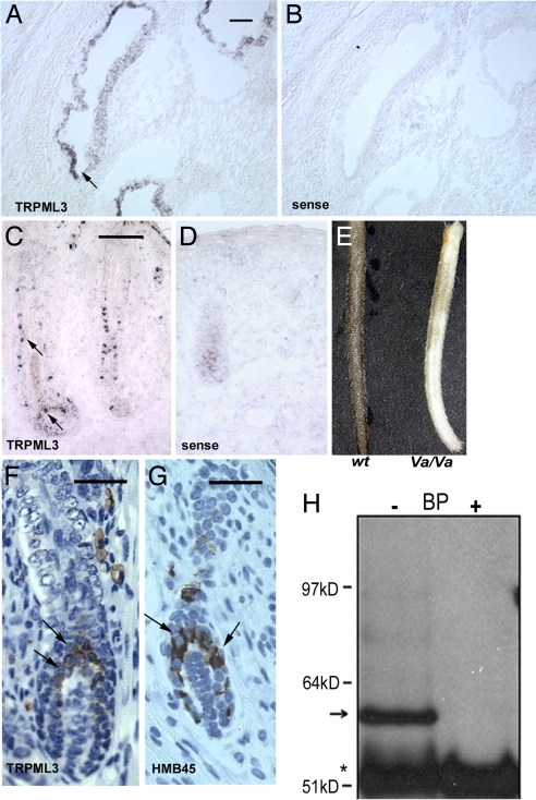
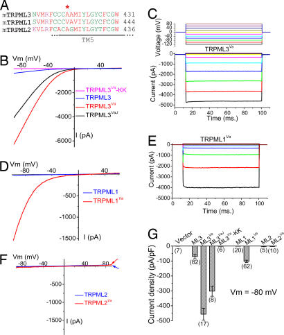
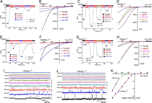
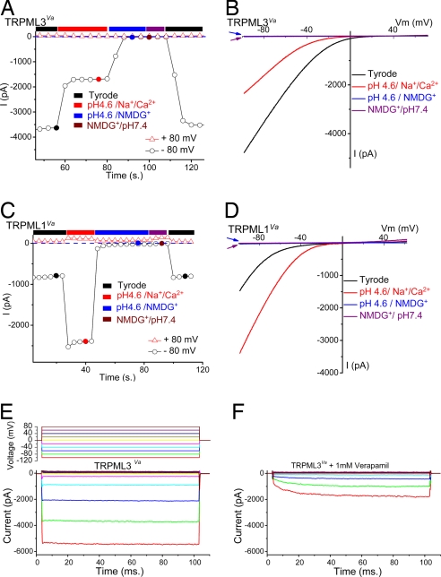
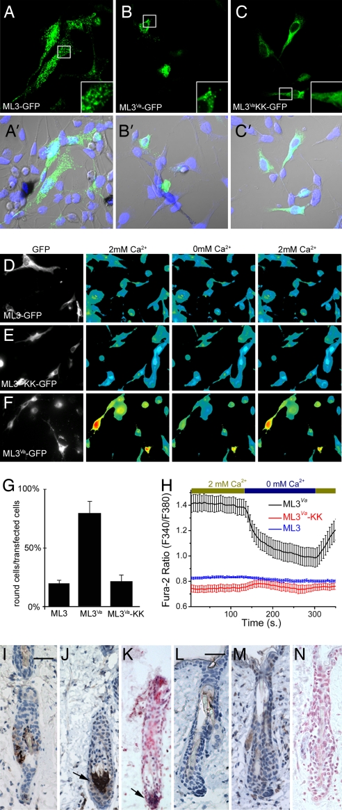
Transient receptor potential (TRP) genes of the mucolipin subfamily (TRPML1-3 and MCOLN1-3) are presumed to encode ion channel proteins of intracellular endosomes and lysosomes. Mutations in human TRPML1 (mucolipin 1/MCOLN1) result in mucolipidosis type IV, a severe inherited neurodegenerative disease associated with defective lysosomal biogenesis and trafficking. A mutation in mouse TRPML3 (A419P; TRPML3(Va)) results in the varitint-waddler (Va) phenotype. Va mice are deaf, exhibit circling behavior due to vestibular defects, and have variegated/dilute coat color as a result of pigmentation defects. Prior electrophysiological studies of presumed TRPML plasma membrane channels are contradictory and inconsistent with known TRP channel properties. Here, we report that the Va mutation produces a gain-of-function that allows TRPML1 and TRPML3 to be measured and identified as inwardly rectifying, proton-impermeant, Ca(2+)-permeant cation channels. TRPML3 is highly expressed in normal melanocytes. Melanocyte markers are lost in the Va mouse, suggesting that their variegated and hypopigmented fur is caused by severe alteration of melanocyte function or cell death. TRPML3(Va) expression in melanocyte cell lines results in high resting Ca(2+) levels, rounded, poorly adherent cells, and loss of membrane integrity. We conclude that the Va phenotype is caused by mutation-induced TRPML3 gain-of-function, resulting in cell death.


Conflict of interest statement

The authors declare no conflict of interest.

Figures

Fig. 1.
Fig. 1.
TRPML3 is highly expressed in the cochlea and melanocytes in skin hair follicles. (A–D) In situ hybridization of mTRPML3 in P0 mice. (B and D) Sense probe. (A) TRPML3 in the stria vascularis of the cochlea. (Scale bar, 100 μm.) (C) TRPML3 expression in a whisker hair follicle. Note the individual cells stained by the antisense probe. (Scale bar, 100 μm.) (E) Skin color differences in WT and Va/Va 6-week-old littermates. (F and G) Immunohistochemical staining with TRPML3-specific antibodies (F) and melanocyte-specific antibodies (HMB45) (G) on paraffin-embedded skin sections of P4 WT mice; antibody staining is brown, and the nuclei are blue. TRPML3-specific staining is visible in the cytoplasm of cells in the upper region and sides of the hair bulb (arrows). (Scale bar, 50 μm.) (G) Melanocytes in similar locations in the hair bulb. (Scale bar, 50 μm.) (H) Immunoprecipitation using anti-TRPML3 antibody was performed in the absence or presence of blocking peptide (BP) from lysates of melan-a2 cells. The TRPML3 band is ≈59 kDa (arrow). The asterisk indicates ab heavy chains; molecular mass markers are shown to the left.
Fig. 2.
Fig. 2.
Large constitutively active inwardly rectifying currents in expressed TRPML1 and TRPML3 Va mutants. (A) Alignment of TRPML (TRPML1, TRPML2, and TRPML3) protein sequences near the Va locus. The red asterisk indicates Ala-419 in TRPML3, which is a proline in Va mice. TM5, putative transmembrane S5. (B) Representative whole-cell currents in HEK293T cells transfected with WT TRPML3, TRPML3Va, TRPLM3VaJ, and TRPML3Va–KK constructs. Currents were elicited by voltage ramps (−100 to +100 mV, 400 ms) with a 4-s interval between ramps in standard extracellular solution (see Materials and Methods). Holding potential (HP) = 0 mV. (C) Large inwardly rectifying whole-cell current elicited by voltage steps (−120 mV to +80 mV, 20 mV increments, step duration = 90 ms) in TRPML3Va-expressing cells. HP = 0 mV. ITRPML3-Va is instantaneously activated at negative potentials. (D) Representative whole-cell currents in TRPML1- and TRPML1Va-expressing HEK293T cells. (E) ITRPML1-Va elicited by voltage steps. (F) No significant current was measured in HEK293T cells transfected with TRPML2 or TRPML2Va. (G) Average inward current densities of TRPML channels at −80 mV normalized by cell capacitance (pF). (n) = number of cells.
Fig. 3.
Fig. 3.
Permeation properties of TRPML1 and TRPML3 channels. (A) TRPML3Va is permeable to both Ca2+ and Mg2+. Currents were initially recorded in external solution and elicited by repeated voltage ramps (−100 to +100 mV, 400 ms) with a 4-s interval between ramps. Data at −80 mV and +80 mV were plotted against time. No significant inward current was seen in the NMDG+ (Na+-free, Ca2+-free) solution. Switching the bath to isotonic (105 mM) Mg2+ or Ca2+ solution yielded smaller, but measurable, current. Divalent ITRPML3-Va currents did not inactivate after repetitive voltage ramps. (B) Whole-cell I–V relations in the presence of isotonic [Ca2+]o and [Mg2+]o. Note the positive reversal potentials (more than +60 mV). (C) Monovalent permeability of ITRPML3-Va in divalent-free (DVF, [Ca2+]i < 10 nM) solution. Large inwardly rectifying currents were elicited by DVF, Na+-containing and DVF, K+-containing solutions and were smaller in DVF Cs+ solution. (D) Representative I–V relations of monovalent Na+, K+, and Cs+ currents in DVF conditions. (E and F) ITRPML1-Va Ca2+ and Mg2+ permeability. Currents elicited by isotonic divalent solutions exhibit weaker voltage dependence than ITRPML1-Va in external solution. Divalent ITRPML1-Va currents did not inactivate over longer durations. (G and H) Large inwardly rectifying monovalent ITRPML1-Va in DVF conditions. At − 80 mV, Na+ ITRPML1-Va in DVF solution was >9-fold larger than the current in external solution. (I) Single-channel currents in an inside-out patch excised from a TRPML3Va-expressing HEK293T cell. Pipette contained external solution; the bath was Cs+-based internal solution (147 mM Cs-Mes). A voltage step [−140 mV to + 80 mV; holding potential (HP) = 0 mV] applied to the inside-out patch elicited channel openings at negative potentials. C and O are closed and open levels, respectively. At negative potentials, substates (S) were frequently observed. (J) Single-channel TRPML1Va currents. (K) TRPML3Va single-channel conductance (−140 mV to −20 mV) was 49 ± 1 pS (n = 4). TRPML1Va single-channel conductance was 76 ± 4 (−140 mV to −100 mV; n = 5) and 11 ± 0.4 pS (−80 mV to −40 mV; n = 5).
Fig. 4.
Fig. 4.
TRPML1 and TRPML3 pH dependence and pharmacological block. (A and B) TRPML3Va is a proton-impermeant cation channel inhibited by low extracellular pH. (A) ITRPML3-Va in external solution was inhibited by ≈60% when the pH of the bath solution was decreased to 4.6. Replacement of permeable cations (Na+/Ca2+/Mg2+/K+) by NMDG+ at pH 4.6 eliminated most inward current. (B) Representative currents in external, pH 4.6, low-pH NMDG+, and pH 7.4 NMDG+ solutions. The amplitudes of NMDG+ currents at pH 7.4 (blue arrow) and pH 4.6 (red arrow) are similar. (C and D) TRPML1Va is a proton-impermeant cation channel strongly enhanced by low extracellular pH. (C) ITRPML1-Va in external solution increased ≈3-fold by acidification to pH 4.6. No significant inward current was seen in NMDG+ solutions (pH 7.4 and pH 4.6). (D) Representative whole-cell currents in pH 4.6 external, low-pH NMDG+, and pH 7.4 NMDG+ solutions. Note that the NMDG+-elicited currents at pH 7.4 (blue arrow) and pH 4.6 (red arrow) are almost indistinguishable. (E) Large inwardly rectifying whole-cell current in a TRPML3Va-expressing cell (protocol, Top) in external solution. (F) Verapamil (1 mM) inhibits and slows activation of currents at negative potentials.
Fig. 5.
Fig. 5.
Elevated [Ca2+]i in TRPML3Va-expressing cells and melanocyte cell death. (A–C) Melan-a2 cells were transfected with GFP-tagged TRPML3 (A and A′), TRPML3Va (B and B′), and TRPML3Va-KK (C and C′) constructs and assayed after 48 h. Note punctate distribution of TRPML3 in melan-a2 cells (A Inset). (B) Round morphology of TRPML3Va-expressing cells. (C and C Inset) Cells expressing the TRPML3Va-KK mutant attach and spread; the fluorescence signal is not punctate. (A′C′) Cells were stained with TO-PRO3 nuclear stain. (D–F) Melan-a2 cells were transfected with GFP-tagged TRPML3 (D), TRPML3Va-KK (E), and TRPML3Va (F) constructs; [Ca2+]i was measured after 20 h. Resting [Ca2+]i in TRPML3- and TRPML3Va-KK-transfected cells was similar to nontransfected cells. In contrast, [Ca2+]i was significantly elevated in TRPML3Va-transfected cells in 2 mM extracellular [Ca2+]. Representative data are from four to eight experiments. (G) Quantification of round vs. spread cells after transfection with the indicated construct (80 ± 9% round cells for TRPML3Va vs. 19 ± 3% round cells for WT TRPML3). The levels of KK mutant were similar to those of WT (19 ± 3% round cells for TRPML3 vs. 22 ± 6% round cells for TRPML3Va-KK transfected cells; n = 3 experiments). (H) In TRPML3Va-transfected cells, Fura-2 ratios in 2 mM [Ca2+]o (1.4 ± 0.06) drop when [Ca2+]o is reduced (nominal 0 mM Ca2+, 1 mM EGTA; 0.99 ± 0.07). [Ca2+]i did not change significantly after removal of extracellular Ca2+ for TRPML3 and TRPML3Va-KK transfected cells (0.82 + 0.01 and 0.75 + 0.03). (I, J, L, and M). Representative immunohistochemical staining, using tyrosinase- (I and L) and TRPML3- (J and M) specific antibodies of paraffin-embedded skin sections from 6-week-old WT (I–K) and Va/Va (L–N) mice. (K and N) Sections were also stained for TRPML3 by in situ hybridization. Antibody staining is brown; nuclei are stained blue. The in situ signal is visible as a blue precipitate with nuclei stained in red. (Scale bar, 50 μm.) (I) Tyrosinase staining of WT hair follicles indicates that melanocytes are primarily in the bulb of the hair follicle. (J) Staining with a TRPML3-specific antibody indicates that TRPML3-positive cells are localized to a similar region in adult mouse hair follicle. (K) TRPML3-specific in situ hybridization; TRPML3 antibody and the in situ probe stain similar regions in the hair follicle bulb. (L and M) Immunohistochemical staining, using tyrosinase- and TRPML3-specific antibodies, was performed side by side with the WT slides. Neither tyrosinase- nor TRPML3- specific signal was detected in the hair bulb of Va/Va mice. In the WT skin sections, ≈26% of the hair follicles were labeled by the tyrosinase antibody (90/347) or by the TRPML3 specific antibody (66/255). No tyrosinase- or TRPML3-specific staining was detected in Va/Va mouse hair follicle bulbs (≈300 hair follicles were analyzed). (N) TRPML3 is absent in hair follicle bulbs from Va/Va skin sections (in situ hybridizations).

References

    1. Pryor PR, Reimann F, Gribble FM, Luzio JP. Traffic. 2006;7:1388–1398. - PMC - PubMed
    1. Venkatachalam K, Hofmann T, Montell C. J Biol Chem. 2006;281:17517–17527. - PMC - PubMed
    1. Vergarajauregui S, Puertollano R. Traffic. 2006;7:337–353. - PMC - PubMed
    1. Song Y, Dayalu R, Matthews SA, Scharenberg AM. Eur J Cell Biol. 2006;85:1253–1264. - PubMed
    1. Di Palma F, Belyantseva IA, Kim HJ, Vogt TF, Kachar B, Noben-Trauth K. Proc Natl Acad Sci USA. 2002;99:14994–14999. - PMC - PubMed

Publication types

Substances