. 2007 Nov 13;104(46):18286-91.
doi: 10.1073/pnas.0708611104. Epub 2007 Nov 7.

Processing of tactile information by the hippocampus

Affiliations

Processing of tactile information by the hippocampus

Antonio Pereira et al. Proc Natl Acad Sci U S A. .

Abstract

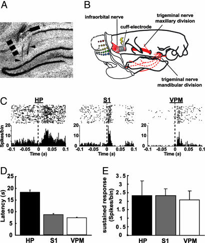
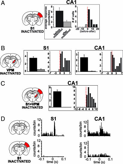
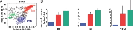
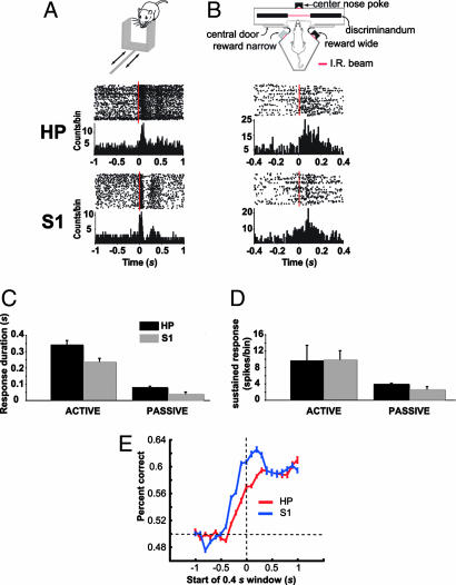
The ability to detect unusual events occurring in the environment is essential for survival. Several studies have pointed to the hippocampus as a key brain structure in novelty detection, a claim substantiated by its wide access to sensory information through the entorhinal cortex and also distinct aspects of its intrinsic circuitry. Novelty detection is implemented by an associative match-mismatch algorithm involving the CA1 and CA3 hippocampal subfields that compares the stream of sensory inputs received by CA1 to the stored representation of spatiotemporal sequences in CA3. In some rodents, including the rat, the highly sensitive facial whiskers are responsible for providing accurate tactile information about nearby objects. Surprisingly, however, not much is known about how inputs from the whiskers reach CA1 and how they are processed therein. Using concurrent multielectrode neuronal recordings and chemical inactivation in behaving rats, we show that trigeminal inputs from the whiskers reach the CA1 region through thalamic and cortical relays associated with discriminative touch. Ensembles of hippocampal neurons also carry precise information about stimulus identity when recorded during performance in an aperture-discrimination task using the whiskers. We also found broad similarities between tactile responses of trigeminal stations and the hippocampus during different vigilance states (wake and sleep). Taken together, our results show that tactile information associated with fine whisker discrimination is readily available to the hippocampus for dynamic updating of spatial maps.


Conflict of interest statement

The authors declare no conflict of interest.

Figures

Fig. 1.
Fig. 1.
Electrically evoked tactile responses in the CA1, S1, and VPM. (A) Localization of electrode tracks in a frontal section of the hippocampus stained with cresyl violet. We used a staggered electrode array targeting both CA1 (arrow) and the dentate gyrus (DG) of the hippocampus (arrowheads), the latter being one of the sources of LFPs for the determination of vigilance state (see Materials and Methods). The dashed line marks the CA1/subiculum border. (B) Schematic diagram showing the location of the cuff electrode implant used to stimulate the IO nerve in behaving rats. (C) Representative single-unit, electrically evoked responses in CA1, S1, and VPM. (Upper) Raster plot of unit spikes, with each line representing a consecutive stimulation trial. (Lower) Summed activity for all trials in 1-ms bins. For data analyses, the initial 2 ms were eliminated to discard electrical stimulus artifacts. HP, hippocampus. (D) Mean ± SEM latencies of electrically evoked responses in CA1, S1, and VPM (n = 3 animals). Response latency to electrical stimulation was estimated as the third peristimulus time histogram consecutive bin, with values falling outside the 95% confidence interval (P < 0.05) of a Poisson distribution fitted to the previous 100-ms interval baseline. (E) Mean ± SEM amplitude of electrically evoked sustained responses in CA1, S1, and VPM (n = 3 animals). This response was calculated by integrating the firing rate over a period of 2–50 ms after the stimulus, subtracted from the average firing rate during the 100-ms preceding the stimulus (15).
Fig. 2.
Fig. 2.
Tactile responses in CA1 depend on the integrity of the somatosensory lemniscal pathway. Muscimol (0.1%) was injected into S1 (A and D), VPM (B), or both (C), and the effects were measured on CA1 units after stimulation of the IO nerve. (A) The left histogram shows muscimol injection caused a great reduction on electrically evoked responses (mean ± SEM) of CA1 units (average of two cases). The right histogram shows the effects of muscimol on sampled units through the difference between average cell activity before and after the injection (red dashed line indicates no difference). (B) Injection of muscimol into the VPM disrupts tactile processing in S1 as expected but also causes a much stronger reduction in CA1 unit activity than inactivation of S1 only (compare with A). (C) Muscimol injection into both S1 and VPM brings about a complete reduction in CA1 unit activity. (D) Peristimulus time histograms of two representative units showing the effects of S1 inactivation on electrically evoked response in S1 and CA1. See Fig. 1 legend for description of peristimulus time histogram plots.
Fig. 3.
Fig. 3.
State dependency of electrically evoked responses in CA1, S1, and VPM. (A) State-space map for one subject (STIM 2) recorded over 24 h (20). Each symbol (filled circle or ×) represents a 1-s epoch of recorded LFPs. The scatter plot is built from two LFP spectral ratios (20). Three distinct clusters corresponding to different states on the wake–sleep cycle are visible: QWK, SWS, and REM sleep. The colored symbols (×) correspond to electrical stimuli delivered to the IO nerve during epochs pertaining to each of these states (blue, QWK; red, SWS; green, REM). (B) Average amplitude (mean ± SEM) of evoked responses of hippocampal, S1, and VPM cells during the three vigilance states in all subjects analyzed. In every recorded area, the electrical stimuli evoked stronger responses when animals were sleeping than during waking. Quantitative analysis of evoked responses is similar to Fig. 1. HP, hippocampus.
Fig. 4.
Fig. 4.
Responses of hippocampal cells to mechanical stimulation of the whiskers in awake and behaving animals. (A) Tactile responses in CA1 and S1 to the passive stimulation of the whiskers. (Top) Schematic of a moving aperture stimulus. The aperture is accelerated across the facial whiskers by a pneumatic solenoid (2). (Middle and Bottom) The response of two representative cells recorded in CA1 and S1, respectively, to the passive stimulation (see Fig. 1 legend for a description of plots). Bin size, 10 ms. The zero time point represents stimulus onset (vertical dashed line). HP, hippocampus. (B) (Top) Schematic of testing chamber used during the active discrimination protocol (see Material and Methods). (Middle and Bottom) Responses of two representative cells in CA1 and S1. Bin size, 10 ms. (C and D) Mean ± SEM response duration and magnitude, respectively, of CA1 and S1 cells evoked during active discrimination and passive whisker stimulation. Amplitude of sustained response was calculated by integrating the firing rate over a period of 300 ms subtracted from the average firing rate during the 100 ms preceding the IR beam breaking. (E) Average LVQ output (one recording session per animal) (n = 5 animals) for CA1 and S1 showing the ability to discriminate between the wide and narrow gap based on the recorded population activity before and after whiskers contacted the aperture. Before the whiskers contacted the aperture, the ability of the LVQ to discriminate between the two apertures was near chance (horizontal dashed line). After the whiskers contacted the aperture, the performance of the LVQ classifier improved above chance for both CA1 and S1 (see Results). The classifier performance for S1 peaked before CA1 (see Results).

References

    1. Nowak RM. Walker's Mammals of the World. Baltimore: Johns Hopkins Univ Press; 1999.
    1. Krupa DJ, Wiest MC, Shuler MG, Laubach M, Nicolelis MA. Science. 2004;304:1989–1992. - PubMed
    1. Krupa DJ, Matell MS, Brisben AJ, Oliveira LM, Nicolelis MA. J Neurosci. 2001;21:5752–5763. - PMC - PubMed
    1. Brecht M, Preilowski B, Merzenich MM. Behav Brain Res. 1997;84:81–97. - PubMed
    1. Carvell GE, Simons DJ. J Neurosci. 1990;10:2638–2648. - PMC - PubMed

Publication types

LinkOut - more resources