Comparative Study
. 2007 Nov 7;27(45):12132-8.
doi: 10.1523/JNEUROSCI.2320-07.2007.

Human motor corpus callosum: topography, somatotopy, and link between microstructure and function

Affiliations
Comparative Study

Human motor corpus callosum: topography, somatotopy, and link between microstructure and function

Mathias Wahl et al. J Neurosci. .

Abstract

The corpus callosum (CC) is the principal white matter fiber bundle connecting neocortical areas of the two hemispheres. Although an object of extensive research, important details about the anatomical and functional organization of the human CC are still largely unknown. Here we focused on the callosal motor fibers (CMFs) that connect the primary motor cortices (M1) of the two hemispheres. Topography and somatotopy of CMFs were explored by using a combined functional magnetic resonance imaging/diffusion tensor imaging fiber-tracking procedure. CMF microstructure was assessed by fractional anisotropy (FA), and CMF functional connectivity between the hand areas of M1 was measured by interhemispheric inhibition using paired-pulse transcranial magnetic stimulation. CMFs mapped onto the posterior body and isthmus of the CC, with hand CMFs running significantly more anteriorly and ventrally than foot CMFs. FA of the hand CMFs but not FA of the foot CMFs correlated linearly with interhemispheric inhibition between the M1 hand areas. Findings demonstrate that CMFs connecting defined body representations of M1 map onto a circumscribed region in the CC in a somatotopically organized manner. The significant and topographically specific positive correlation between FA and interhemispheric inhibition strongly suggests that microstructure can be directly linked to functional connectivity. This provides a novel way of exploring human brain function that may allow prediction of functional connectivity from variability of microstructure in healthy individuals, and potentially, abnormality of functional connectivity in neurological or psychiatric patients.


Figures

Figure 1.
Figure 1.
Combined fMRI/DTI procedure to track CMFs. a, Tracked fibers after first tracking step: a large ROI (blue rectangle) was set that covered the fMRI-defined M1 face, hand, and foot representations of both hemispheres. b, A second ROI (blue rectangle) was placed within the area of the CC identified by the first tracking step, and fiber tracking was performed again. c, A target ROI (blue rectangle) was centered on the knob of the right precentral gyrus, an accepted anatomical landmark for the M1 hand representation (Yousry et al., 1997). The closely adjacent fMRI-defined M1 hand area is depicted in green. d, Only those tracked fibers projecting into this target ROI were retained. Note that, because of the tracking procedure that was started bidirectionally from the seeds in the CC, fibers are retained also in a homologous region of the hand area of the left M1. A similar procedure was performed for the fMRI-defined M1 lip (magenta in a, b) and foot areas (orange; tracked fibers not shown). e, Selected voxels for FA calculation (purple) superimposed on the tracked hand CMFs (green), axial view at high magnification. f, Data from the same subject as in e, midsagittal view. R, Right.
Figure 2.
Figure 2.
a, fMRI-defined M1 representations (lip: light red; hand: light green; foot: yellow) and tracked CMFs (lip: dark red; hand: dark green; foot: orange) visualized as 3D objects in one subject. b, The CMFs (midsagittal view) pass through the posterior body and isthmus of the CC. Note the somatotopic order with lip CMFs located most ventrally and anteriorly, the foot CMFs located most dorsally and posteriorly, and the hand CMFs located in between. R, Right.
Figure 3.
Figure 3.
Midsagittal topography of hand CMFs (green) and foot CMFs (orange) in all 12 subjects rendered in a modified Talairach space. The blue contours indicate the outlines, if the hand and foot CMFs from all subjects were superimposed on one plot. Note consistent location of the CMFs in the posterior body and isthmus of the CC in all subjects, and ventral and/or anterior location of the hand CMFs relative to the foot CMFs in all subjects except subject 7 and 9.
Figure 4.
Figure 4.
a, b, Superimposition of the hand CMFs (a) and foot CMFs (b) of all subjects onto one CC in modified Talairach space. Color bars indicate the number of subjects with CMFs at the same site. c, The topographical extent of the superimposed hand CMFs (green) and foot CMFs. Note the separation of hand and foot CMFs along a ventral/anterior-to-dorsal/posterior axis.
Figure 5.
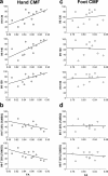
Figure 5.
Linkage of microstructure with functional connectivity of the human motor CC. a, b, Linear regression analysis revealed a positive correlation between the FA of the hand CMFs and the magnitude of IHI between the hand areas of M1 at intensities of the conditioning pulse of 130% RMT (r = 0.59; p = 0.04), 140% RMT (r = 0.66; p = 0.02), and 150% RMT (r = 0.59; p = 0.04) (a) and a negative correlation with the threshold intensity (IHI-T) of the conditioning pulse [given as a percentage of maximum stimulator output (%MSO)] to produce 25% IHI (r = −0.60; p = 0.04) and 50% IHI (r = −0.64; p = 0.02) (b). Each point reflects FA and IHI from one individual subject. c, d, This correlation was absent between the FA of the foot CMFs and the IHI (c) and IHI threshold (d) between the hand areas of M1 (p values >0.6 at all intensities of the conditioning pulse).

Comment in

References

    1. Aboitiz F, Scheibel AB, Fisher RS, Zaidel E. Fiber composition of the human corpus callosum. Brain Res. 1992;598:143–153. - PubMed
    1. Andres FG, Mima T, Schulman AE, Dichgans J, Hallett M, Gerloff C. Functional coupling of human cortical sensorimotor areas during bimanual skill acquisition. Brain. 1999;122:855–870. - PubMed
    1. Basser PJ, Pajevic S, Pierpaoli C, Duda J, Aldroubi A. In vivo fiber tractography using DT-MRI data. Magn Reson Med. 2000;44:625–632. - PubMed
    1. Beaulieu C. The basis of anisotropic water diffusion in the nervous system—a technical review. NMR Biomed. 2002;15:435–455. - PubMed
    1. Behrens TE, Berg HJ, Jbabdi S, Rushworth MF, Woolrich MW. Probabilistic diffusion tractography with multiple fiber orientations: what can we gain? NeuroImage. 2007;34:144–155. - PMC - PubMed

Publication types

LinkOut - more resources