Comparative Study
. 2007 Nov 7;27(45):12147-55.
doi: 10.1523/JNEUROSCI.3655-07.2007.

Dual modes of Munc18-1/SNARE interactions are coupled by functionally critical binding to syntaxin-1 N terminus

Affiliations
Comparative Study

Dual modes of Munc18-1/SNARE interactions are coupled by functionally critical binding to syntaxin-1 N terminus

Mikhail Khvotchev et al. J Neurosci. .

Abstract

The SM (Sec1/Munc18-like) protein Munc18-1 and the soluble N-ethylmaleimide-sensitive factor attachment protein (SNAP) receptor (SNARE) proteins syntaxin-1, SNAP-25, and synaptobrevin/VAMP (vesicle-associated membrane protein) constitute the core fusion machinery for synaptic vesicle exocytosis. Strikingly, Munc18-1 interacts with neuronal SNARE proteins in two distinct modes (i.e., with isolated syntaxin-1 alone in a "closed" conformation and with assembled SNARE complexes containing syntaxin-1 in an "open" conformation). However, it is unclear whether the two modes of Munc18/SNARE interactions are linked. We now show that both Munc18/SNARE interaction modes involve the same low-affinity binding of the extreme syntaxin-1 N terminus to Munc18-1, suggesting that this binding connects the two Munc18/SNARE interaction modes to each other. Using transfected cells as an in vitro assay system, we demonstrate that truncated syntaxins lacking a transmembrane region universally block exocytosis, but only if they contain a free intact N terminus. This block is enhanced by coexpression of either Munc18-1 or SNAP-25, suggesting that truncated syntaxins block exocytosis by forming an untethered inhibitory SNARE complex/Munc18-1 assembly in which the N-terminal syntaxin/Munc18 interaction is essential. Introduction of an N-terminal syntaxin peptide that disrupts this assembly blocks neurotransmitter release in the calyx of Held synapse, whereas a mutant peptide that does not disrupt the SNARE complex/Munc18 assembly has no effect. Viewed together, our data indicate that binding of Munc18 to the syntaxin N terminus unites different modes of Munc18/SNARE interactions and is essential for exocytic membrane fusion.


Figures
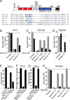
Figure 1.
Figure 1.
Syntaxin-mediated inhibition of regulated and constitutive exocytosis involves Munc18-1 and SNAP-25 binding. A, Domain structure of syntaxin-1. Amino acid residues are numbered on top and on the right of the alignment. The conserved N-terminal sequences of syntaxin-1 to -5 (Synt1A–5A) are aligned below, with shared residues highlighted in blue. Residues critical for the syntaxin-5A/Sly1 interaction and corresponding residues in other syntaxins are shown in red. The D3 position (D5 for syntaxin-5A) is marked with an asterisk. TMR, Transmembrane region. B, C, Munc18-1 potentiates the inhibition of exocytosis by syntaxin-1A fragments [which act by blocking fusion at the plasma membrane (Khvotchev and Sudhof, 2004)]. Data shown are assays of regulated exocytosis in PC12 cells (stimulated with 56 mm K+ for 15 min (B) and of constitutive exocytosis in HEK293 cells (C). Cells were cotransfected with an hGH reporter vector and various expression vectors as indicated; exocytosis was measured 72 h (B, F) or 48 h (C–E, G, H) after transfection. Regulated exocytosis in PC12 cells is shown as the difference in release of hGH (calculated as the fraction of total hGH produced) between stimulated and control cells. Constitutive hGH release is displayed as the ratio of extracellular-to-intracellular hGH for a given sample. D, Sly1 relieves the inhibition of hGH secretion by syntaxin-5A, which blocks the secretory pathway in the Golgi complex. E, The inhibitory activity of syntaxin-1A1–243 is reversed by mutations that impair stable SNARE complex formation but not Munc18-1 binding (L205D and I209D) (Matos et al., 2003) (see supplemental Fig. 3, available at www.jneurosci.org as supplemental material). F, G, The isolated SNARE motif of syntaxin-1A is unable to inhibit regulated (F) or constitutive (G) exocytosis. Syntaxin-1A1–243 and syntaxin-1A173–243 were C-terminally fused to EYFP (to stabilize expression). Both EYFP-fusion proteins form SDS-resistant SNARE complexes in cells (see supplemental Fig. 4, available at www.jneurosci.org as supplemental material). H, SNAP-25 potentiates the inhibition of exocytosis mediated by syntaxin-1A fragments similar to Munc18-1. Error bars indicate SEM.
Figure 2.
Figure 2.
Free intact syntaxin-1A (Synt1A) N terminus is required for the inhibition of exocytosis. A, B, Inactivation of the inhibitory activity of syntaxin-1A1–243 by addition of an N-terminal myc-tag. To ensure that the apparent effect of the myc-tag is not attributable to a loss of expression, we measured the relative inhibition of exocytosis as a function of increasing amounts of transfected plasmid (B). All samples contained equal amounts of total transfected DNA (1.05 μg/well), with the remainder made up of empty expression vector. C, Mutations in, or truncation of, the N terminus of syntaxin-1A inactivate the inhibition of constitutive exocytosis by syntaxin-1A1–243. All data shown are from a representative experiment performed in duplicate and independently repeated at least twice with comparable results. Error bars indicate SEM.
Figure 3.
Figure 3.
Effects of plasma membrane syntaxins 2–4 and Golgi syntaxin-5A on constitutive exocytosis also depends on N-terminal sequences. Secretion assays were performed as described in Figure 1. Syntaxin (Synt)-2, -3, -4, and -5A fragments were modeled after the syntaxin-1A1–243 fragment based on protein sequence alignment. A, Effects of individual syntaxins and their combinations. B, Comparison of effects of syntaxins with or without the D3R mutation (D5R for syntaxin-5A). C, D, Unequal contributions of syntaxins to constitutive exocytosis inhibition by introducing D3R mutations in various combinations of syntaxins-2 and -4 (C) and syntaxins-3 and -4 (D). All data shown are from a representative experiment performed in duplicate and independently repeated at least twice with comparable results. Error bars indicate SEM. WT, Wild type.
Figure 4.
Figure 4.
FRET reveals essential role of the N-terminal syntaxin-1A sequence in Munc18-1 binding. A, Venus-tagged syntaxin-1A1–243 exhibits efficient FRET when bound to Munc18-1 containing monomeric Cerulean inserted at position 94 but not at position 24. Fluorescence emission spectra (excitation, 433 nm) were collected from HEK293 cells transfected with expression vectors encoding Venus-tagged syntaxin-1A1–243 and Cerulean-tagged Munc18-1. B, Intact N terminus of syntaxin-1A (Synt1A) is required for efficient FRET with Munc18-1. HEK293 cells were transfected with Munc18-1 C94, Venus GFP, or various syntaxin-1A fragments fused to Venus. Cell extracts were mixed so that each sample contained an equal amount of Cerulean (excitation, 433 nm; emission, 475 nm) and Venus (excitation, 515 nm; emission, 528 nm) fluorescence. After incubation to allow formation of Munc18-1/syntaxin-1A complexes, fluorescence emission spectra (excitation, 433 nm) were collected. Data are from a single experiment repeated multiple times with similar results.
Figure 5.
Figure 5.
N-terminal sequence of syntaxin-1A binds to Munc18-1: analysis by NMR. A, Superposition of 1H-15N HSQC spectra of 15N-labeled syntaxin-1A2–243 (0.2 mm) in the absence (black) or presence (red) of a 1.5 molar excess of unlabeled full-length Munc18-1. The spectra are plotted at high contour levels to help visualizing the sharp cross-peaks from flexible residues. B, C, Superposition of 1H-15N HSQC spectra of 15N-labeled syntaxin-1A2–180 (0.05 mm) in the absence (black) or presence (red) of 1.5 molar excess of unlabeled Munc18-1 N-terminal domain (residues 1–136); C displays an expansion of the spectra in B (denoted by the dashed rectangle) to illustrate the shifts and/or broadening of selected cross-peaks of syntaxin-1A (labeled with the corresponding assignments) after binding to the Munc18-1 N-terminal domain. In A and B, the cross-peaks corresponding to T5 and T10 from the N-terminal motif of syntaxin-1A are labeled. D, Structure of the syntaxin-1A Habc domain (residues 27–146; orange) and Munc18-1 N-terminal domain (residues 1–136; blue) within the crystal structure of the Munc18-1/syntaxin-1A complex (Misura et al., 2000). The residues of the Habc domain that are substantially shifted and/or broadened by binding to the Munc18-1 N-terminal domain are shown in green. The N- and C-terminal residues of both domains are labeled. The dashed line illustrates that the N-terminal sequence of syntaxin-1A, which was not visible in the crystal structure of the Munc18-1/syntaxin-1A complex, could easily reach the binding site observed for the N-terminal sequence of Sed5p on the Sly1p N-terminal domain in the crystal structure of the Sed5p/Sly1p complex (Bracher and Weissenhorn, 2002).
Figure 6.
Figure 6.
Wild-type (WT) but not mutant (D3R) N-terminal syntaxin-1A peptide interferes with Munc18-1/SNARE complex assembly. A, 1D 13C-edited 1H-NMR spectra of 2 μm 13C-labeled Munc18-1 in the absence or presence of 5 μm unlabeled preassembled SNARE complex containing syntaxin-1A2–253, Synaptobrevin 21–96, and the two SNARE motifs of SNAP-25 (residues 11–82 and 141–203), before and after addition of 480 μm wild-type or D3R mutant syntaxin-1A N-terminal peptides (residues 2–16 with addition of a C-terminal cysteine residue). B, SDS-PAGE analysis of protein complexes used in NMR experiments shown in A. The asterisk indicates a proteolytic fragment of Munc18-1; such proteolysis occurs in a flexible loop and does not impair binding to syntaxin-1A (Misura et al., 2000) or the SNARE complex (our unpublished results).
Figure 7.
Figure 7.
Effect of N-terminal syntaxin-1A peptides on synaptic vesicle exocytosis in the calyx of Held synapse. A, B, Sample traces of calcium current and capacitance measurements at the presynaptic terminal of calyx of Held performed after dialysis of 0.5 mm wild-type syntaxin-1A peptide (A) and 0.5 mm D3R syntaxin-1A peptide (B). C, Dose-dependent rundown of exocytosis after dialysis of wild-type or D3R syntaxin-1A peptides. WT, Wild type; n, number of independent experiments.

References

    1. Arac D, Murphy T, Rizo J. Facile detection of protein-protein interactions by one-dimensional NMR spectroscopy. Biochemistry. 2003;42:2774–2780. - PubMed
    1. Arac D, Dulubova I, Pei J, Huryeva I, Grishin NV, Rizo J. Three-dimensional structure of the rSly1 N-terminal domain reveals a conformational change induced by binding to syntaxin 5. J Mol Biol. 2005;346:589–601. - PubMed
    1. Borst JG, Helmchen F, Sakmann B. Pre- and postsynaptic whole-cell recordings in the medial nucleus of the trapezoid body of the rat. J Physiol (Lond) 1995;489:825–840. - PMC - PubMed
    1. Bracher A, Weissenhorn W. Crystal structures of neuronal squid Sec1 implicate inter-domain hinge movement in the release of t-SNAREs. J Mol Biol. 2001;306:7–13. - PubMed
    1. Bracher A, Weissenhorn W. Structural basis for the Golgi membrane recruitment of Sly1p by Sed5p. EMBO J. 2002;21:6114–6124. - PMC - PubMed

Publication types

LinkOut - more resources