Comparative Study
. 2007 Nov 7;27(45):12156-67.
doi: 10.1523/JNEUROSCI.1898-07.2007.

The neuropeptide VGF produces antidepressant-like behavioral effects and enhances proliferation in the hippocampus

Affiliations
Comparative Study

The neuropeptide VGF produces antidepressant-like behavioral effects and enhances proliferation in the hippocampus

Smita Thakker-Varia et al. J Neurosci. .

Abstract

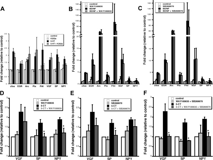
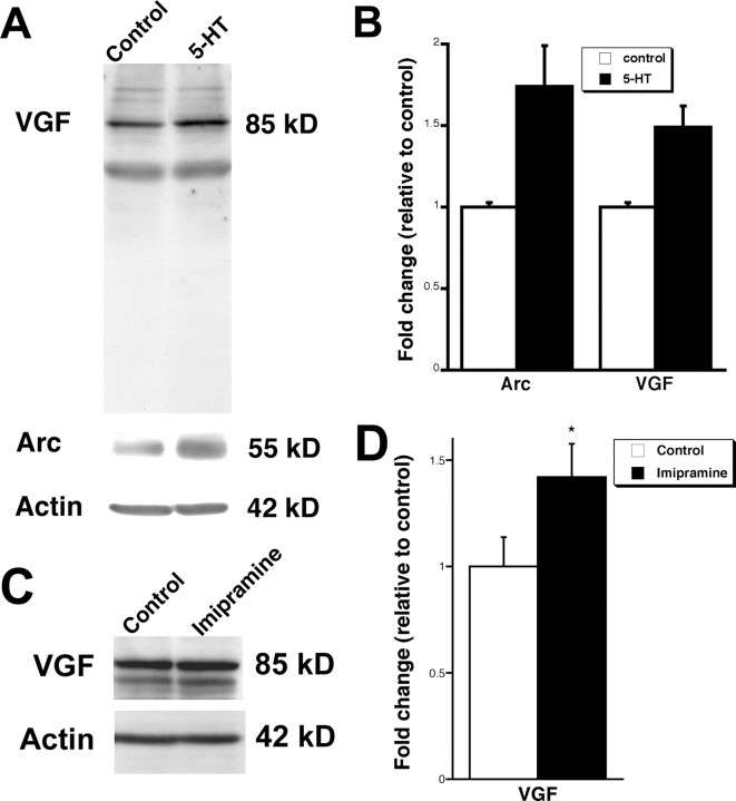
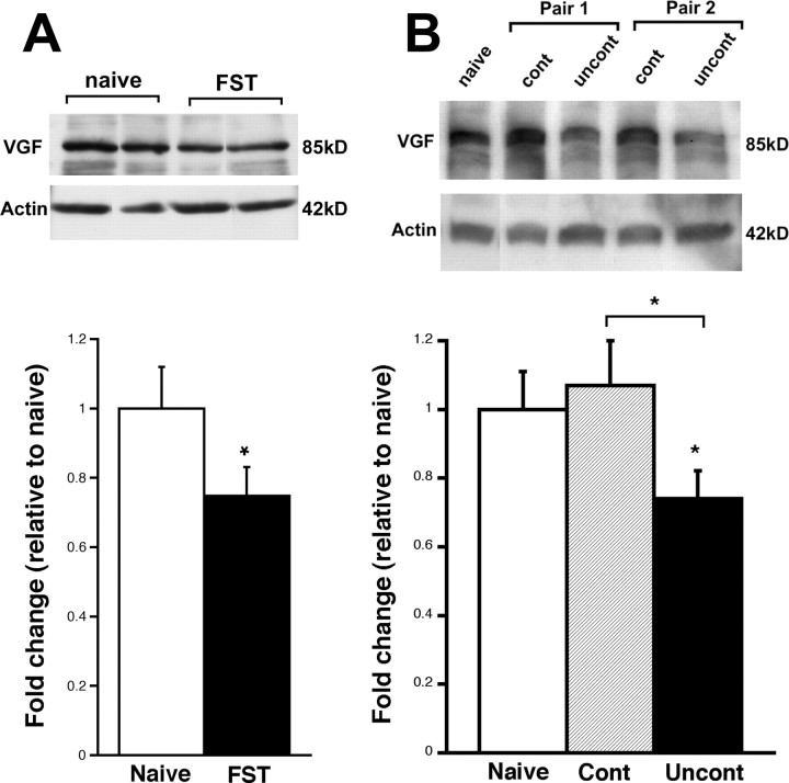
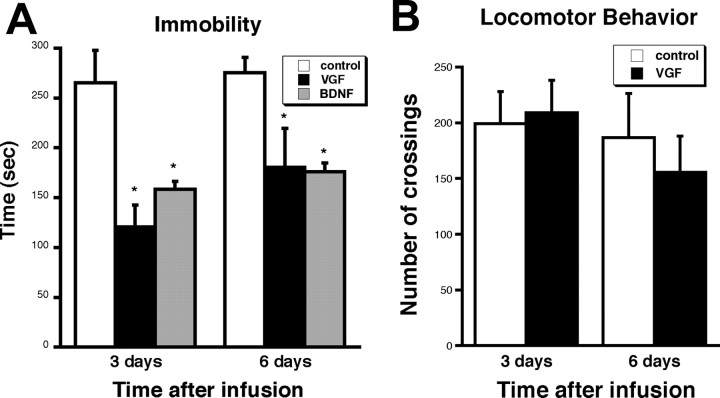
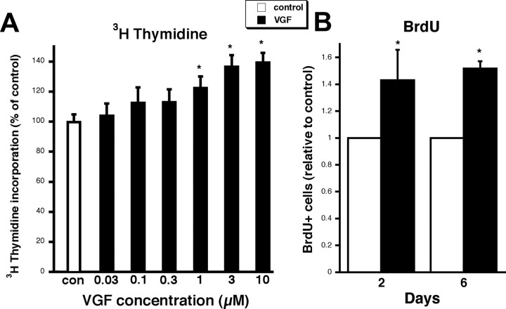
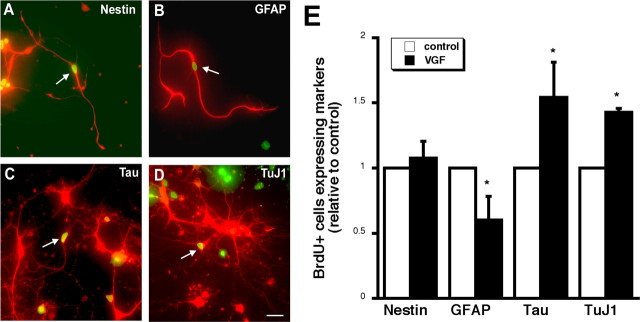
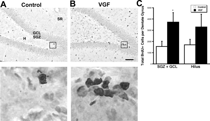
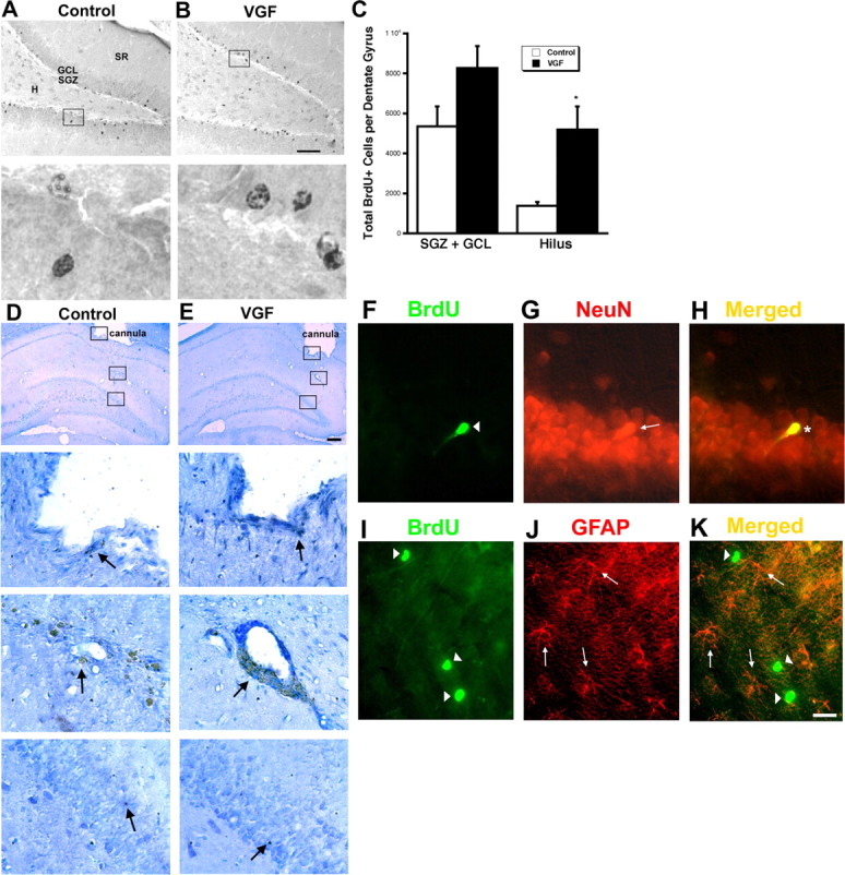
Brain-derived neurotrophic factor (BDNF) is upregulated in the hippocampus by antidepressant treatments, and BDNF produces antidepressant-like effects in behavioral models of depression. In our previous work, we identified genes induced by BDNF and defined their specific roles in hippocampal neuronal development and plasticity. To identify genes downstream of BDNF that may play roles in psychiatric disorders, we examined a subset of BDNF-induced genes also regulated by 5-HT (serotonin), which includes the neuropeptide VGF (nonacronymic). To explore the function of VGF in depression, we first investigated the expression of the neuropeptide in animal models of depression. VGF was downregulated in the hippocampus after both the learned helplessness and forced swim test (FST) paradigms. Conversely, VGF infusion in the hippocampus of mice subjected to FST reduced the time spent immobile for up to 6 d, thus demonstrating a novel role for VGF as an antidepressant-like agent. Recent evidence indicates that chronic treatment of rodents with antidepressants increases neurogenesis in the adult dentate gyrus and that neurogenesis is required for the behavioral effects of antidepressants. Our studies using [(3)H]thymidine and bromodeoxyuridine as markers of DNA synthesis indicate that chronic VGF treatment enhances proliferation of hippocampal progenitor cells both in vitro and in vivo with survival up to 21 d. By double immunocytochemical analysis of hippocampal neurons, we demonstrate that VGF increases the number of dividing cells that express neuronal markers in vitro. Thus, VGF may act downstream of BDNF and exert its effects as an antidepressant-like agent by enhancing neurogenesis in the hippocampus.


Figures

Figure 1.
Figure 1.
5-HT regulates transcription independent of BDNF and requires differential activation of either 5-HT1 and/or 5-HT7. A, Average gene expression in hippocampal cells preincubated with the trk receptor antagonist K252a (200 nm) for 30 min before 5-HT treatment (1 μm) followed by real time RT-PCR for a panel of genes. Only one gene, PAI, showed a significantly lower response to 5-HT plus K252a than 5-HT alone. *5-HT plus K252a is significantly different from 5-HT alone (p < 0.05, ANOVA; n = 4). B, C, Average gene expression in hippocampal cells preincubated with 5-HT receptor antagonists for 30 min before BDNF treatment (50 ng/ml) followed by real-time RT-PCR for a panel of genes. B, 5-HT1A antagonist (WAY100635; 10 μm). C, 5-HT7 receptor antagonist (SB269970; 10 μm). No statistical difference was detected between BDNF plus receptor antagonists and BDNF alone for any of the genes (p > 0.05, ANOVA; n = 3). D–F, Average gene expression in neurons preincubated with inhibitors to 5-HT receptor subtypes before 5-CT (1 μm) treatment followed by real-time RT-PCR for the neuropeptides VGF, SP, and NPY. D, 5-HT1A antagonist (WAY100635; 10 μm). E, 5-HT7 antagonist (SB269970; 10 μm). F, Combination of WAY100635 plus SB269970. One group of cells in each experiment was treated with inhibitor alone and showed no significant change in expression relative to vehicle control. *Significant reduction of transcription in the presence of inhibitor plus 5-CT relative to 5-CT alone (p < 0.05, ANOVA; n = 3). All samples were first normalized to GAPDH and then represented as an average ratio of vehicle controls ± SE.
Figure 2.
Figure 2.
5-HT and imipramine upregulate Arc and VGF protein levels. A, Western blot analysis of Arc and VGF expression after 5-HT (1 μm) exposure of hippocampal cultures for 6 h. B, Quantitation of protein levels indicates that Arc and VGF are upregulated. The bars represent average protein expression ± SE (n = 2). Data are first normalized to actin to control for protein loading, and data are expressed as a fold of expression in vehicle controls. C, Representative Western blot of VGF expression in the hippocampus after intraperitoneal injection of imipramine (10 mg/kg) for 21 d. D, Quantitation of protein levels indicates VGF is upregulated. The bars represent average protein expression ± SE (n = 4, 6). Data are first normalized to actin, which controls for protein loading, and data are expressed as a fold of expression in vehicle-injected rats. *p < 0.05, unpaired t test.
Figure 3.
Figure 3.
VGF expression is decreased in two models of depression. A, Representative Western blot of VGF protein expression in naive rodents and those subjected to FST. Each lane represents one animal. All data are normalized to actin expression. The bars represent average VGF protein levels relative to naive ± SE (n = 4, 5). *p < 0.05, unpaired t test. B, Western blot of VGF protein expression in a representative naive animal and two representative pairs of animals subjected to controllable or uncontrollable stress. All data are normalized to actin expression. The bars represent average VGF protein expression ± SE in rodents subjected to controllable stress or uncontrollable stress (n = 7) relative to naive rats (n = 3). *p < 0.05, ANOVA.
Figure 4.
Figure 4.
VGF intrahippocampal infusions decrease immobility in FST. A, Mice were injected bilaterally with saline, VGF (100 ng), or BDNF (500 ng) and scored on FST (6 min) performed 3 and 6 d postsurgery. The bars represent average time spent immobile for all animals ± SE (n = 5). *p < 0.05, t test. B, Mice were also scored for locomotor behavior in the open field test. The bars represent the average number of quadrant crossings ± SE in a 15 min time period scored 3 and 6 d after surgery (n = 5). *p < 0.05, t test.
Figure 5.
Figure 5.
VGF enhances DNA synthesis in hippocampal cultures. A, VGF increases [3H]thymidine incorporation in hippocampal cultures in a dose-dependent manner. Hippocampal neurons were treated with VGF in concentrations ranging from 0.03 to 10 μm or vehicle control from time of plating to 48 h. Cells were then loaded with 1 μCi/ml [3H]thymidine at 37°C for 4 h. Data are expressed as mean percentage of control ± SE (n = 8). *p < 0.05, ANOVA. B, Hippocampal cells were treated with VGF (3 μm) or vehicle control at time of plating. BrdU was added to cells at 48 h and cells were fixed 2 or 6 d later. The bars represent fraction of total cells that were BrdU+ expressed as an average fold change relative to control ± SE. Approximately 200–700 total cells were counted per dish, and there were four dishes per condition per experiment. Each bar represents a cell count from three independent experiments (∼2400–8400 total cells). *p < 0.05, ANOVA.
Figure 6.
Figure 6.
VGF promotes the differentiation of neuronal cells. A–D, BrdU and cell-specific markers colocalize after 6 d of differentiation. Freshly plated hippocampal cells were treated with 3 μm VGF or vehicle control at time of plating. BrdU was added to cultures at 48 h, and cells were fixed 6 d later. The arrows indicate double-labeled cells with a BrdU+ nucleus (green) and nestin (A), GFAP (B), Tau (C), and TuJ1 (red) (D). Scale bar, 50 μm. E, Quantitation of BrdU+ cells expressing cell-specific markers after 6 d of differentiation in vitro. Data represent fraction of BrdU+ cells coexpressing cell-specific markers including the precursor markers nestin and GFAP and the mature neuronal markers Tau and TuJ1 (∼50 BrdU+ cells/dish; n = 3). *p < 0.05, ANOVA.
Figure 7.
Figure 7.
Intrahippocampal infusion of VGF increases BrdU+ cells in the SGZ plus GCL. A, B, Saline (A) or VGF (4 μg) (B) was infused daily via a cannula into the hippocampi of adult male rats for 7 d followed by a single BrdU injection (100 mg/kg). The animals were killed 2 h later. BrdU+ cells (black) are visible in the subdivisions of the dentate gyrus including the hilus (H), subgranular zone (SGZ), granule cell layer (GCL), and stratum radiatum (SR) in coronal sections. Scale bar, 80 μm. Enlarged images of boxed regions are shown below. C, Quantitation of total BrdU+ cells in the various regions of the dentate gyrus. The bars represent average BrdU+ cells ± SE (n = 7, 9). *p < 0.05, t test.
Figure 8.
Figure 8.
VGF-induced BrdU+ cells survive to 21 d and differentiate primarily into neurons. A–C, VGF-induced BrdU+ cells survive to 21 d. A, B, Saline (A) or VGF (4 μg) (B) was infused daily via a cannula into the hippocampi of adult male rats for 7 d followed by a single BrdU injection (100 mg/kg). The animals were killed 21 d later. BrdU+ cells (black) are visible in the subdivisions of the dentate gyrus including the hilus (H), subgranular zone (SGZ), granule cell layer (GCL), and stratum radiatum (SR) in coronal sections. Scale bar, 80 μm. Enlarged images of boxed regions are shown below. C, Quantitation of total BrdU+ cells in the various regions of the dentate gyrus. The bars represent average BrdU+ cells ± SE (n = 8). *p < 0.05, t test. D, E, Repeated infusions of saline or VGF do not cause damage to the dentate gyrus. Saline (D) or VGF (4 μg) (E) was infused daily via a cannula into the hippocampi of adult male rats for 7 d. The animals were killed 21 d later and processed for cleaved caspase-3 immunohistochemistry. Location of guide cannula is shown at low magnification (arrowhead). Scale bar, 150 μm. Enlarged images of boxed regions showing tip of cannula, site of infusion, and dentate gyrus, respectively, are shown below. Caspase-3-positive cells are indicated by arrows. F–H, BrdU+ cells that survive primarily express neuronal markers. F, BrdU+ cells (green; arrowhead). G, NeuN+ cells (red; arrow). H, Merged image showing colocalization (yellow; asterisk). I–K, BrdU+ cells that survive do not express glia markers. I, BrdU+ cells (green; arrowheads). J, GFAP+ cells (red; arrows). K, Merged image showing no double-labeled cells (yellow). Scale bar, 20 μm.

References

    1. Abad MA, Enguita M, DeGregorio-Rocasolano N, Ferrer I, Trullas R. Neuronal pentraxin 1 contributes to the neuronal damage evoked by amyloid-β and is overexpressed in dystrophic neurites in Alzheimer's brain. J Neurosci. 2006;26:12735–12747. - PMC - PubMed
    1. Aberg MA, Aberg ND, Hedbacker H, Oscarsson J, Eriksson PS. Peripheral infusion of IGF-I selectively induces neurogenesis in the adult rat hippocampus. J Neurosci. 2000;20:2896–2903. - PMC - PubMed
    1. Adell A, Castro E, Celada P, Bortolozzi A, Pazos A, Artigas F. Strategies for producing faster acting antidepressants. Drug Discov Today. 2005;10:578–585. - PubMed
    1. Alder J, Thakker-Varia S, Bangasser DA, Kuroiwa M, Plummer MR, Shors TJ, Black IB. Brain-derived neurotrophic factor-induced gene expression reveals novel actions of VGF in hippocampal synaptic plasticity. J Neurosci. 2003;23:10800–10808. - PMC - PubMed
    1. Alder J, Thakker-Varia S, Crozier RA, Shaheen A, Plummer MR, Black IB. Early presynaptic and late postsynaptic components contribute independently to brain-derived neurotrophic factor-induced synaptic plasticity. J Neurosci. 2005;25:3080–3085. - PMC - PubMed

Publication types

MeSH terms