Comparative Study
. 2007 Nov 7;27(45):12413-8.
doi: 10.1523/JNEUROSCI.0719-07.2007.

Loss-of-function of human PINK1 results in mitochondrial pathology and can be rescued by parkin

Affiliations
Comparative Study

Loss-of-function of human PINK1 results in mitochondrial pathology and can be rescued by parkin

Nicole Exner et al. J Neurosci. .

Abstract

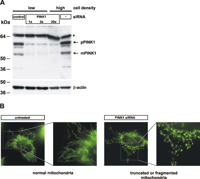
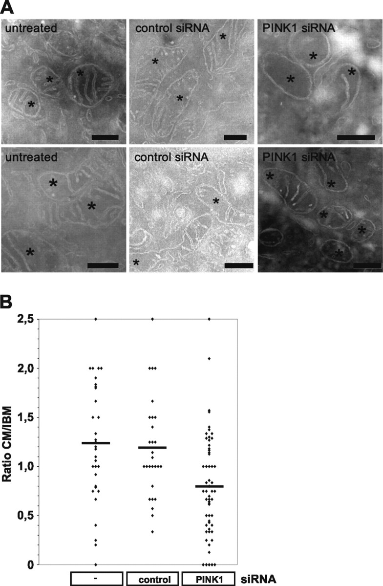
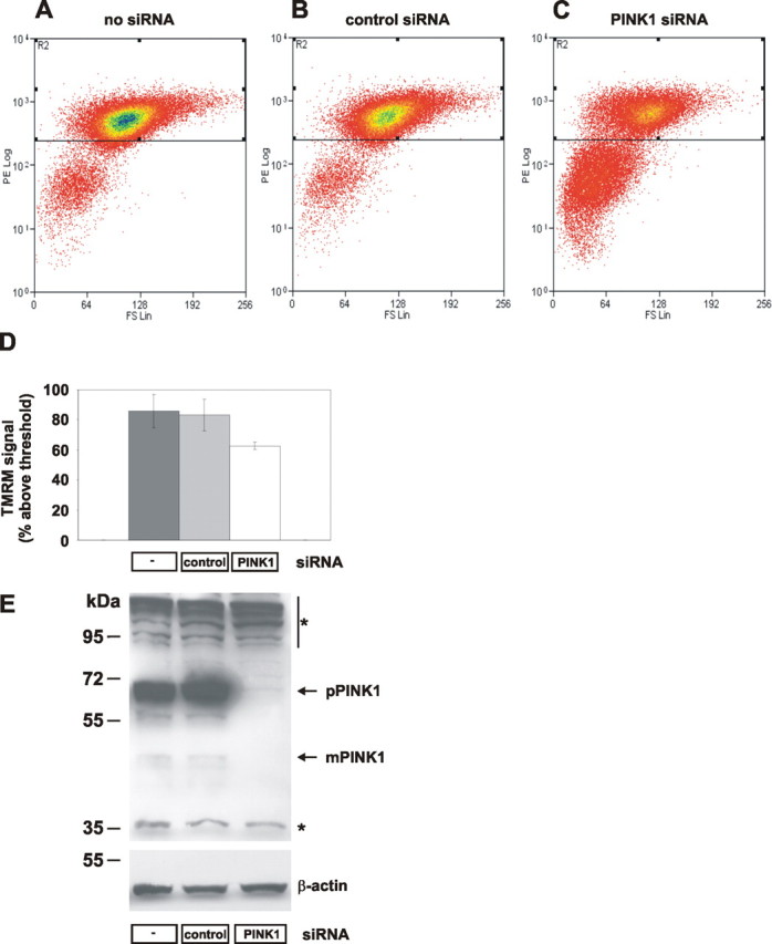
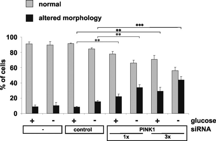
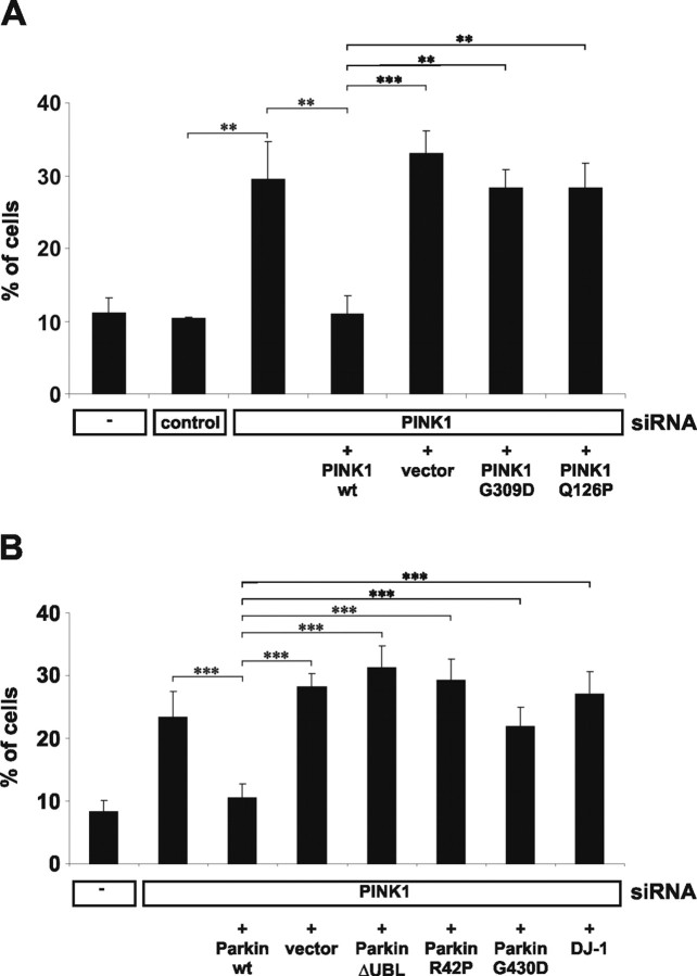
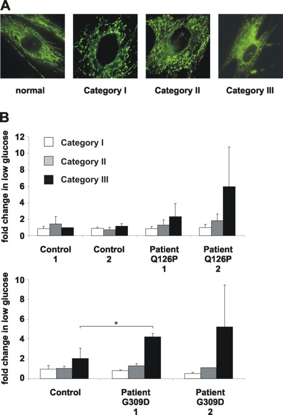
Degeneration of dopaminergic neurons in the substantia nigra is characteristic for Parkinson's disease (PD), the second most common neurodegenerative disorder. Mitochondrial dysfunction is believed to contribute to the etiology of PD. Although most cases are sporadic, recent evidence points to a number of genes involved in familial variants of PD. Among them, a loss-of-function of phosphatase and tensin homolog-induced kinase 1 (PINK1; PARK6) is associated with rare cases of autosomal recessive parkinsonism. In HeLa cells, RNA interference-mediated downregulation of PINK1 results in abnormal mitochondrial morphology and altered membrane potential. Morphological changes of mitochondria can be rescued by expression of wild-type PINK1 but not by PD-associated PINK1 mutants. Moreover, primary cells derived from patients with two different PINK1 mutants showed a similar defect in mitochondrial morphology. Human parkin but not PD-associated mutants could rescue mitochondrial pathology in human cells like wild-type PINK1. Our results may therefore suggest that PINK1 deficiency in humans results in mitochondrial abnormalities associated with cellular stress, a pathological phenotype, which can be ameliorated by enhanced expression of parkin.


Figures

Figure 1.
Figure 1.
RNA silencing of PINK1 causes alteration in mitochondrial morphology in human cells. A, siRNA-mediated downregulation of PINK1. Untransfected HeLa cells (−), or cells transfected with a control siRNA (control), or an siRNA against PINK1 were analyzed for endogenous PINK1 protein levels. Cells were transfected at two different cell densities with 5, 15, or 100 pmol of siRNA (1×, 3×, or 20×) (pPINK1, precursor form; mPINK1, mature form). A cross-reactive protein is marked by an asterisk. B, Representative examples of normal or altered (truncated or fragmented) mitochondrial morphologies including higher magnifications are shown.
Figure 2.
Figure 2.
Altered cristae morphology after siRNA-mediated downregulation of PINK1. A, Cells were fixed, cryosectioned, and analyzed by standard electron transmission microscopy. Representative mitochondrial sections are shown for cells untreated (left), treated with control siRNA (middle), and treated with PINK1 siRNA (right). Asterisks indicate sections through mitochondria. Scale bars, 300 nm. B, Quantitative analysis of CM and IBM length.
Figure 3.
Figure 3.
Loss of PINK1 affects the mitochondrial membrane potential. A–C, HeLa cells were transfected on 3 consecutive days with no siRNA (A), 10 pmol of control siRNA (B), or 10 pmol of PINK1 siRNA (C) and stained with TMRM. Cells were separated by FACS according to size (forward scatter, x-axis) and red TMRM fluorescence (y-axis). Cells with intact mitochondrial membrane potential had TMRM fluorescence >102; below are cells with loss of membrane potential. D, Mean percentage of cells with intact membrane potential. Error bars indicate SE. E, Effects of siRNA treatments on PINK1 expression were determined by Western blot for endogenous PINK1 protein (top; pPINK1, precursor form; mPINK1, mature form) and control β-actin (bottom). Cross-reactive proteins are marked by asterisks.
Figure 4.
Figure 4.
Glucose deprivation increases changes in mitochondrial morphology. Untransfected HeLa cells or cells transfected with siRNA as indicated were investigated for mitochondrial morphology by life imaging. Cells with normal mitochondria (gray bars) or altered mitochondria (black bars) were counted after incubation of cells in standard cell culture medium (high glucose; +) or in low-glucose (−) medium. Results represent mean values of three independent experiments with error bars indicating SD (ANOVA; **p ≤ 0.01; ***p ≤ 0.001).
Figure 5.
Figure 5.
Rescue of the morphological alterations caused by reduced PINK1 expression. A, Cells were cotransfected with siRNA and either empty vector or PINK1 wild type (wt), PINKG309D, PINK1Q126P siRNA-resistant constructs as indicated. Note the rescue of mitochondrial pathology by wt PINK1 but not by mutant PINK1. B, Cells were cotransfected with PINK1 siRNA, wild-type (wt) parkin; the parkin mutations ΔUBL, R42P, and G430D; as well as wt DJ-1. Note the rescue of mitochondrial pathology by wt parkin, but not by DJ-1. (ANOVA; **p ≤ 0.01; ***p ≤ 0.001). Error bars indicate SD.
Figure 6.
Figure 6.
Truncation and fragmentation of mitochondria in fibroblasts from patients with PINK1 mutations. A, Mitochondrial fragmentation phenotypes with increasing severity (category I, swollen; category II, truncated and swollen; category III, fragmented) as observed in fibroblasts of PARK6 patients. B, Fibroblasts of controls or PD patients carrying the PINK1Q126P or PINK1G309D mutations were analyzed for mitochondrial morphology after a shift to low-glucose medium. Changes in the percentage of cells in each category are represented by white (category I), gray (category II), and black (category III) bars (ANOVA, *p ≤ 0.05). Error bars indicate SD.

Similar articles

Cited by

References

    1. Baba M, Nakajo S, Tu PH, Tomita T, Nakaya K, Lee VM, Trojanowski JQ, Iwatsubo T. Aggregation of alpha-synuclein in Lewy bodies of sporadic Parkinson's disease and dementia with Lewy bodies. Am J Pathol. 1998;152:879–884. - PMC - PubMed
    1. Beilina A, Van Der Brug M, Ahmad R, Kesavapany S, Miller DW, Petsko GA, Cookson MR. Mutations in PTEN-induced putative kinase 1 associated with recessive parkinsonism have differential effects on protein stability. Proc Natl Acad Sci USA. 2005;102:5703–5708. - PMC - PubMed
    1. Bonifati V, Rizzu P, van Baren MJ, Schaap O, Breedveld GJ, Krieger E, Dekker MC, Squitieri F, Ibanez P, Joosse M, van Dongen JW, Vanacore N, van Swieten JC, Brice A, Meco G, van Duijn CM, Oostra BA, Heutink P. Mutations in the DJ-1 gene associated with autosomal recessive early-onset parkinsonism. Science. 2003;299:256–259. - PubMed
    1. Chen L, Feany MB. Alpha-synuclein phosphorylation controls neurotoxicity and inclusion formation in a Drosophila model of Parkinson disease. Nat Neurosci. 2005;8:657–663. - PubMed
    1. Clark IE, Dodson MW, Jiang C, Cao JH, Huh JR, Seol JH, Yoo SJ, Hay BA, Guo M. Drosophila pink1 is required for mitochondrial function and interacts genetically with parkin. Nature. 2006;441:1162–1166. - PubMed

Publication types

MeSH terms