. 2007 Dec 12;26(24):5083-92.
doi: 10.1038/sj.emboj.7601922. Epub 2007 Nov 8.

RAP-1 and the RAL-1/exocyst pathway coordinate hypodermal cell organization in Caenorhabditis elegans

Affiliations

RAP-1 and the RAL-1/exocyst pathway coordinate hypodermal cell organization in Caenorhabditis elegans

Ester W Frische et al. EMBO J. .

Abstract

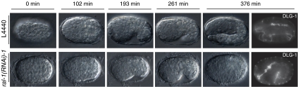
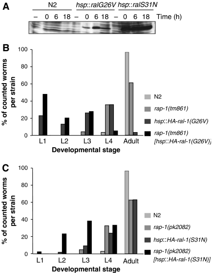
The small Ras-like GTPase Rap1 has been identified as a regulator of integrin activation and cadherin-mediated cell-cell contacts. Surprisingly, null mutants of RAP-1 in Caenorhabditis elegans are viable and fertile. In a synthetic lethal RNAi screen with C. elegans rap-1 mutants, the Ras-like GTPase ral-1 emerged as one of seven genes specifically required for viability. Depletion of exoc-8 and sec-5, encoding two putative RAL-1 effectors and members of the exocyst complex, also caused lethality of rap-1 mutants, but did not affect wild-type worms. The RAP-1 and the RAL-1/exocyst pathway appear to coordinate hypodermal cell movement and elongation during embryonic development. They mediate their effect in part through targeting the alpha-catenin homologue HMP-1 to the lateral membrane. Genetic interactions show that the RAP-1 and RAL-1/exocyst pathway also act in parallel during larval stages. Together these data provide in vivo evidence for the exocyst complex as a downstream RAL-1 effector in cell migration.


Figures

Figure 1
Figure 1
Schematic representation of the genomic organization of the rap-1 locus and detection of mutant alleles. Filled boxes indicate exons and the line underneath the graphic representation indicates the position of the deletion in rap-1(tm861) animals and the star indicates the amber codon (amino acid 130) in the pk2082 allele. Numbers indicate the position of the rap-1 locus on the C27B7 cosmid. Below are PCR reactions shown, performed on single-worm lysates that demonstrate the presence of the wild-type allele in +/+ and +/− worms and the absence of the wild-type allele in −/− worms. An arrowhead indicates a 170 bp Sau3A product that is characteristic for the pk2082 allele. Arrows indicate the position of the PCR primers used for detection of the deletion and wild-type alleles.
Figure 2
Figure 2
Phenotype of rap-1 mutant embryos carrying the dlg-1∷GFP gene subjected to control, ral-1, sec-5 or exoc-8(RNAi). Phenotypes of rap-1 mutant embryos carrying the dlg-1∷GFP marker (FZ271) that were derived from animals subjected to RNAi for ral-1, sec-5 or exoc-8. DLG-1 indicates dlg-1∷GFP expression, AJM-1 indicates immunofluorescence staining using the MH27 antibody. Genes used for RNAi are indicated above each panel. L4440 is the empty vector control RNAi. (AH) show class I embryos, (IL) class II embryos. (A–D) and (I–J) show focal planes to visualize hypodermal cells, (E–H) and (K–L) are focal planes at the level of the gut. The most anterior and posterior visible seam cells are marked with *. In all cases, AJM staining was performed (only shown for class II, subjected to ral-1 (RNAi)), which demonstrated clear colocalization with DLG-1∷GFP). Class I embryos are characterized by halted migration of hypodermal cells as examplified by a ral-1(RNAi) embryo (arrow in B) or misalignment of hypodermal cells as shown here for a sec-5 and exoc-8(RNAi) embryo (C, D). Halted migration can be discerned from early stages of migration by comparison of the developmental stage of the gut. Note that during normal hypodermal cell migration (A, E), the gut has not yet extended along the entire length of the embryos, in contrast to the gut of the ral-1(RNAi) embryo, which has established clear adherens junctions and shows a more mature pharynx (B, F). In some cases, a gap between intestinal and pharyngeal cells is observed (arrowhead H). In class II embryos, hypodermal cells do not envelope the entire embryo (arrow I, J) and a clearly recognizable gut structure is found on the outside of the embryo (arrowhead K). All embryos are oriented such that their pharynx is on the left side. Scale bar: 10 μm.
Figure 3
Figure 3
Phenotype of rap-1 mutant embryos carrying the SCM∷GFP gene subjected to control, ral-1, sec-5 or exoc-8(RNAi) (A–D respectively). Expression of the seam cell marker SCM∷GFP in nuclei of the disorganized hypodermis of class II embryos. Hypodermal cell borders are visualized using the AJM-1 antibody (red). Only one focal plane is shown per condition, seam nuclei are also present in other slices (data not shown). Scale bar: 10 μm.
Figure 4
Figure 4
Localization of HMP-1 in wild-type or rap-1 mutant embryos subjected to control, ral-1, sec-5 or exoc-8(RNAi). dlg-1∷GFP and HMP-1 staining in a lateral view of wild type carrying dlg-1∷GFP (FZ224) (A, B, E, F, I, J, M, N) and rap-1 mutant embryos (FZ271) (C, D, G, H, K, L, O, P), derived from animals subjected to either control L4440 (A–D), ral-1(RNAi) (E–H), sec-5(RNAi) (I–L) and exoc-8(RNAi) (M–P). Pictures show hypodermal cells in which DLG-1 indicates dlg-1∷GFP expression and HMP-1 indicates immunofluorescence staining using the P1E11 antibody. The most anterior and posterior visible seam cells are marked with *. Scale bar: 10 μm.
Figure 5
Figure 5
Time-lapse analysis of rap-1 mutant embryos subjected to control and ral-1(RNAi). Time lapse recordings of embryos derived from dlg-1∷GFP;rap-1(tm861) mutant (FZ271) embryos on control L4440 (upper panels) or ral-1(RNAi) (lower panels). Last timepoint also shows DLG-1∷GFP expression in the gut of the same embryo.
Figure 6
Figure 6
Schematic representation of the genomic organization of the sec-5 locus. Filled boxes indicate exons and the line underneath the graphic representation indicates the position of the deletion in sec-5(tm1443) animals and the star indicates the stop codon (amino acid 389) in the pk2357 and pk2358 allele. Numbers indicate the position of the rap-1 locus on the T23G7 cosmid. Arrows indicate the position of the PCR primers used for detection of the deletion and wild-type alleles.
Figure 7
Figure 7
Effect of heat-shock-driven expression of RAL-1 proteins in wild-type and rap-1 mutant backgrounds. (A) Western blot of total lysates from L1 larvae. Larvae were lysed without heat shock (−) or at 0 h (0), 6 h (6) or 18 h (18) after a 2-h heat shock. Probing was done with the 12CA5 antibody, which recognizes the HA-tag at the N-terminus of overexpressed proteins. (B, C) Embryos subjected to heat shock were allowed to develop 5 days before determination of developmental stage. Percentage of animals at the different developmental stages of the total number of hatched animals is shown for overexpression RAL-1(G26V) and RAL-1(S31N) in (B) and (C), respectively.

References

    1. Andrews R, Ahringer J (2007) Asymmetry of early endosome distribution in C. elegans embryos. PLoS ONE 2: e493. - PMC - PubMed
    1. Asha H, de Ruiter ND, Wang MG, Hariharan IK (1999) The Rap1 GTPase functions as a regulator of morphogenesis in vivo. EMBO J 18: 605–615 - PMC - PubMed
    1. Bos JL (2005) Linking Rap to cell adhesion. Curr Opin Cell Biol 17: 123–128 - PubMed
    1. Bossinger O, Klebes A, Segbert C, Theres C, Knust E (2001) Zonula adherens formation in Caenorhabditis elegans requires dlg-1, the homologue of the Drosophila gene discs large. Dev Biol 230: 29–42 - PubMed
    1. Bryant DM, Stow JL (2004) The ins and outs of E-cadherin trafficking. Trends Cell Biol 14: 427–434 - PubMed

Publication types

MeSH terms

Substances

LinkOut - more resources