. 2007 Oct 31;1(1):e2.
doi: 10.1371/journal.pntd.0000002.

Development of highly organized lymphoid structures in Buruli ulcer lesions after treatment with rifampicin and streptomycin

Affiliations

Development of highly organized lymphoid structures in Buruli ulcer lesions after treatment with rifampicin and streptomycin

Daniela Schütte et al. PLoS Negl Trop Dis. .

Abstract

Background: Buruli ulcer caused by Mycobacterium ulcerans is an infection of the subcutaneous tissue leading to chronic necrotising skin ulcers. The pathogenesis is associated with the cytocidal and immunosuppressive activities of a macrolide toxin. Histopathological hallmark of progressing disease is a poor inflammatory response despite of clusters of extracellular bacilli. While traditionally wide excision of the infected tissue was the standard treatment, provisional WHO guidelines now recommend an eight week pre-treatment with streptomycin and rifampicin.

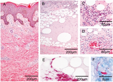
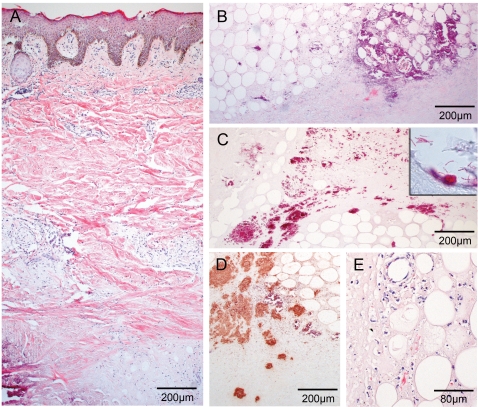
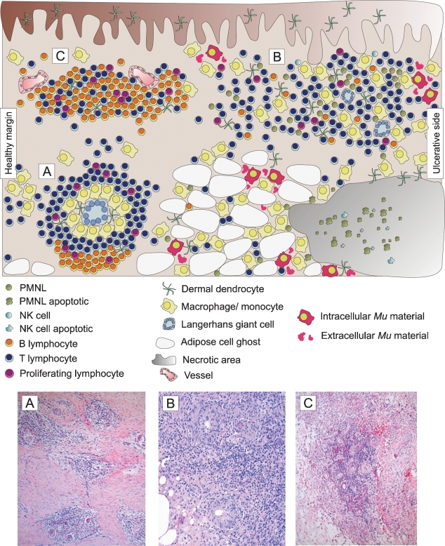
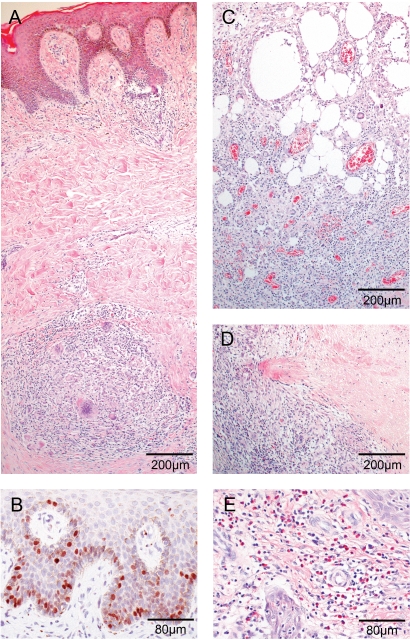
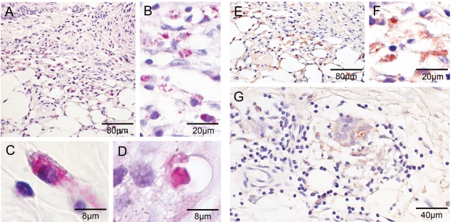
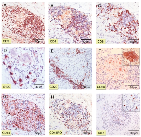
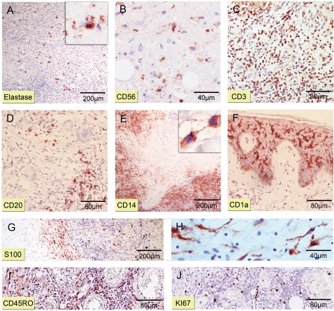
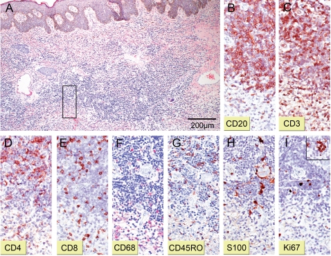
Methodology/principal findings: We conducted a detailed immunohistochemical analysis of tissue samples from Buruli patients who received antibiotic treatment. Cellular immune response along with bacterial load and distribution were monitored. We demonstrate that this treatment leads to the development of highly organized cellular infiltration surrounding areas of coagulative necrosis. Diffuse infiltrates, granulomas and dense lymphocyte aggregation close to vessels were observed. Mycobacterial material was primarily located inside mononuclear phagocytes and microcolonies consisting of extracellular rod-shaped mycobacteria were no longer found. In observational studies some patients showed no clinical response to antibiotic treatment. Corresponding to that, one of five lesions analysed presented with huge clusters of rod-shaped bacilli but no signs of infiltration.

Conclusions/significance: Results signify that eight weeks of antibiotic treatment reverses local immunosuppression and leads to an active inflammatory process in different compartments of the skin. Structured leukocyte infiltrates with unique signatures indicative for healing processes developed at the margins of the lesions. It remains to be analysed whether antibiotic resistance of certain strains of M. ulcerans, lacking patient compliance or poor drug quality are responsible for the absent clinical responses in some patients. In future, analysis of local immune responses could serve as a suitable surrogate marker for the efficacy of alternative treatment strategies.


Conflict of interest statement

The authors have declared that no competing interests exist.

Figures

Figure 1
Figure 1. Histopathological characteristics associated with untreated Buruli ulcer lesions.
Histological sections of specimen from untreated Buruli ulcer lesions stained with HE (A to D) and ZN (E, F), respectively. Photographs are taken at magnification ×40 (A, B, E), ×100 (C, D) or ×1000 (F). (A) Vasculitis associated minor leukocyte infiltration around vessels with intact dermal connective tissue and epidermal hyperplasia. (B) Extensive areas of necrosis in the deeper dermis and large fat cell ghosts with slight leukocyte infiltrates. (C) Slight cellular infiltration mainly composed of PMNL. (D) Ongoing necrotic/apoptotic processes surrounding a focus of mycobacterial microcolonies. (E) Typical clusters of extracellular bacteria between adipose cell ghosts. (F) Sometimes also single bacilli can be spotted inside necrotic regions.
Figure 2
Figure 2. One patient exhibits strong histopathological signs for a progressive Buruli ulcer and large mycobacterial clumps.
Histological sections of the non-responding patient stained with HE (A, B, D) and ZN (C) or polyclonal anti-leprae antibody (pAbLep; E). Magnification ×40 (A, B, C, E) and ×100 (D). (A) Tissue shows typical signs of advanced Buruli ulcer as deep dermal necrosis, calcification or epidermal hyperplasia. Leucocytes are found in rare cases around vessels and hardly ever between fat ghosts. (B) Adipose tissue and its surroundings are highly necrotic and happen to accumulate fat ghosts and calcification. (C) The centre of the necrotic lesion harbours tremendous clumps of rod-shaped mycobacteria. (D) Staining with pAbLep demonstrates the focal clusters of live bacteria in the necrotic core. (E) If cellular infiltration occurs in small amounts within some parts of the intradermal adipose tissue cells display apoptotic features.
Figure 3
Figure 3. Three major types of cellular infiltration can be distinguished in antibiotic treated BU patients.
Upper part: Schematic overview of cellular infiltration patterns and distribution of mycobacterial material. Lower part: Three types of cellular infiltration are documented with HE. (A) Granuloma formation in the connective tissue; magnification ×40. (B) Diffuse heterogeneous cellular infiltration of the connective and adipose tissue; magnification ×100. (C) Follicle-like lymphocyte focus adjacent to vessels; magnification ×40.
Figure 4
Figure 4. Histopathology of four patients in response to rifampicin/streptomycin bi-therapy.
Histological sections representative for four patients stained with HE (A, C, D, E) and polyclonal antibody against proliferation marker Ki67 (B). Magnification ×40 (A, C, E) and ×100 (B, E). (A) Psoriatic epidermal hyperplasia typically seen in Buruli. Diffuse mixed cellular infiltrates in upper and granuloma formation with Langhans' giant cells lymphocytes in deeper dermis. (B) Ki67 staining reveals elevated proliferation levels of keratinocytes in epidermal basal layer. (C) Cell ghosts of the adipose tissue characteristic for Buruli infection. Massive mixed cellular infiltrates between fat ghosts mainly consisting of macrophages/monocytes and formation of new blood vessels. (D) Necrotic area in deep tissue encircled by extensive cellular infiltrates. (E) Focal eosinophilia found at margins of the excised area distant to ulcerative centre.
Figure 5
Figure 5. Bacterial load and distribution in four patients.
Histological sections representative for four patients stained with ZN (A to D) and polyclonal anti-leprae antibody (pAbLep; E to G) to demonstrate distribution of mycobacterial material in the excised lesion. Counterstain was performed with haematoxylin and pictures taken at magnifications of ×100 (A, E), ×200 (G), ×400 (B, F) and ×1000 (C, D). (A, E) Serial sections demonstrate equal detection sensitivity with ZN and pAbLep. Higher amounts of bacterial material near the ulcerative edges with macrophages being the most prevalent leucocytes. (B, F) Close-up of A and E, respectively. Foci of rounded bacteria located extra- and intracellular between fat ghosts. (C) Macrophage presenting with mycobacterial material within phagosomal vesicles. (D) Mycobacterial residues appear in rare cases inside Langhans' giant cell phagosomal vacuoles. (G) Small granuloma with giant cell formation and traces of mycobacteria inside a phagosome.
Figure 6
Figure 6. Detailed organization of granulomas.
Histological serial sections representative for four patients were stained with antibodies against different cellular surface or cytoplasmic markers (counterstain haematoxylin). Magnification ×40 (I), ×100 (A, B, C, E, F, G, H, Iinsert), ×200 (D) and ×400 (Finsert). (A, B, C) Staining with CD3, CD4 and CD8, respectively, reveals a belt of helper as well as cytotoxic T lymphocytes surrounding the APC core. (D) S100+ dermal dendrocytes (dDC) spread among T lymphocytes in the outer layer of a granuloma. (E) Focus of CD20+ B lymphocytes at the border of a granuloma. (F) CD68+ APC in the centre of a representative granuloma; insert shows large Langhans' giant cell. (G) Remarkable large amounts of membrane bound (arrow) and soluble (arrowhead) CD14 can be observed. (H) Distribution of activated CD45RO+ lymphocytes. (I) Proliferating Ki67+ cells indicate the active status of granulomas.
Figure 7
Figure 7. Composite of mixed cellular infiltrates.
Histological sections representative for four patients stained with antibodies against different cellular markers (counterstain haematoxylin). Magnifications at ×40 (A, E, G), ×100 (C, D, F, I, J), ×200 (Ainsert, B, H) or ×1000 (Einsert). (A) Only few scattered PMNL staining positive for Elastase were found within cellular infiltrates. (Ainsert, B) Neutrophilic and NK cell (CD56+) foci inside necrotic areas display signs of advanced apoptosis. (C) Lymphocytes mainly expose a CD3+ phenotype. (D) Small focal CD20+ lymphocyte spots are scattered through infiltrates. (E) Staining against CD14 illustrates large numbers of histiocytes enclosing necrotic tissue shedding massive amounts of sCD14 (insert). (F) Levels of epidermal CD1a+ Langerhans cells are remarkably elevated compared to healthy skin. (G) Aggregation of S100+ dDCs near necrotic spots. (H) Elongated cellular appendices of dDCs reach into the necrotic tissue. (I) Large numbers of lymphocytes are CD45RO+. (J) Same area as in (I). Proliferating lymphocytes are highly Ki67+.
Figure 8
Figure 8. Follicle-like lymphocyte foci reveal organized cellular formation.
Serial sections of a representative lymphocyte focus stained with HE (A; magnification ×40) or with antibodies against different cellular markers and haematoxylin as counterstain (B to I; magnification ×100). (A) Large aggregate of lymphocytes in the upper dermal layer between two venules. Square marks area of magnification chosen for pictures B to I. (B) CD20+ B lymphocytes build the most abundant cell subset. (C, D, E) Staining against CD3, CD4 and CD8, respectively, exposes also high loads of T lymphocytes with T helper clearly being more prevalent than cytotoxic T cells. (F) APCs (CD68+) are scattered throughout the entire structure. (G) Activated lymphocytes appear positive for CD45RO staining. (H) S100+ dermal dendrocytes (dDC) lie in between lymphocytes. (I) Ki67 staining demonstrates certain proliferation of cells and occasional appearance of discrete spots of hyperproliferation (insert).

References

    1. Asiedu A, Scherpbier RW, Raviglione M. Geneva, Switzerland: World Health Organization; 2000. Buruli ulcer, Mycobacterium ulcer infection.
    1. Johnson PD, Stinear TP, Small PL, Pluschke G, Merritt RW, et al. Buruli ulcer (M. ulcerans infection): new insights, new hope for disease control. PLoS Med. 2005;2:e108. - PMC - PubMed
    1. George KM, Chatterjee D, Gunawardana G, Welty D, Hayman J, et al. Mycolactone: a polyketide toxin from Mycobacterium ulcerans required for virulence. Science. 1999;283:854–857. - PubMed
    1. Stinear TP, Mve-Obiang A, Small PL, Frigui W, Pryor MJ, et al. Giant plasmid-encoded polyketide synthases produce the macrolide toxin of Mycobacterium ulcerans. Proc Natl Acad Sci USA. 2004;101:1345–1349. - PMC - PubMed
    1. Snyder D, Small PL. Uptake and cellular actions of mycolactone, a virulence determinant for Mycobacterium ulcerans. Microb Pathog. 2003;34:91–101. - PubMed

Publication types