. 2008 Apr;117(2):123-35.
doi: 10.1007/s00412-007-0131-7. Epub 2007 Nov 8.

Persistence of DNA threads in human anaphase cells suggests late completion of sister chromatid decatenation

Affiliations

Persistence of DNA threads in human anaphase cells suggests late completion of sister chromatid decatenation

Lily Hui-Ching Wang et al. Chromosoma. 2008 Apr.

Abstract

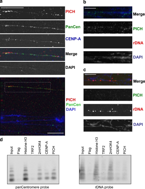
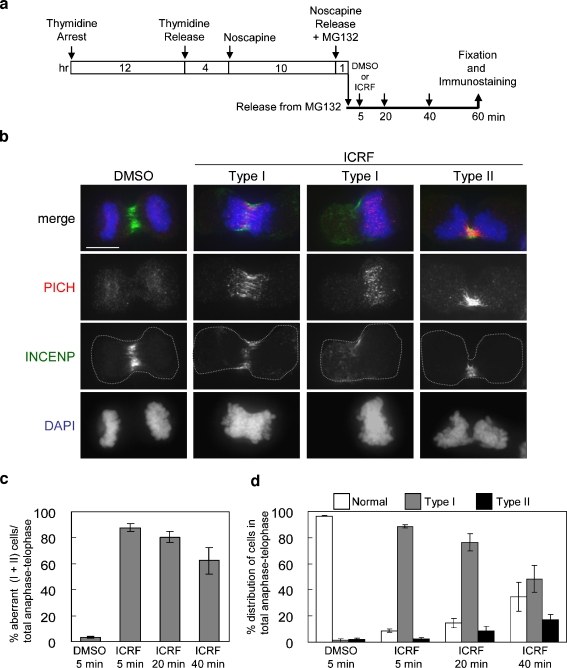
PICH (Plk1-interacting checkpoint helicase) was recently identified as an essential component of the spindle assembly checkpoint and shown to localize to kinetochores, inner centromeres, and thin threads connecting separating chromosomes even during anaphase. In this paper, we have used immuno-fiber fluorescence in situ hybridization and chromatin-immunoprecipitation to demonstrate that PICH associates with centromeric chromatin during anaphase. Furthermore, by careful analysis of PICH-positive anaphase threads through FISH as well as bromo-deoxyurdine and CREST labeling, we strengthen the evidence that these threads comprise mainly alphoid centromere deoxyribonucleic acid. Finally, by timing the addition of ICRF-193 (a specific inhibitor of topoisomerase-II alpha) to cells synchronized in anaphase, we demonstrate that topoisomerase activity is required specifically to resolve PICH-positive threads during anaphase (as opposed to being required to prevent the formation of such threads during earlier cell cycle stages). These data indicate that PICH associates with centromeres during anaphase and that most PICH-positive threads evolve from inner centromeres as these stretch in response to tension. Moreover, they show that topoisomerase activity is required during anaphase for the resolution of PICH-positive threads, implying that the complete separation of sister chromatids occurs later than previously assumed.


Figures

Fig. 1
Fig. 1
Immuno-fiber staining showing PICH associates with centromeric chromatin fibers in anaphase. Extended chromatin fibers were prepared from either anaphase (30-min release from Noscapine, ad, and right panel in e) or metaphase cells (5 min release from Noscapine, left panel in e) and then examined for colocalization of PICH with the indicated markers. All bars indicate 5 μm. a PICH associates with BrdU-positive anaphase DNA fibers. b A chromatin fiber showing positivity for PICH, histone H3 dimethylated at lysine 4 (2mH3K4), and CENP-A. c A PICH-positive chromatin fiber showing staining with histone H3 trimethylated at lysine 9 (3mH3K9) and CENP-A. d Chromatin fibers stained for PICH and histone H3 acetylated at lysine 18 (AcH3K18): PICH-positive thread shows no AcH3K18 staining (left) and vice versa; AcH3K18-positive fiber shows no PICH decoration (right). e INCENP associated with PICH in metaphase chromatin fibers (left) but dissociated from PICH-positive fibers in anaphase (right). f Quantification results showing the positivity of indicated markers on PICH-positive chromatin fibers
Fig. 2
Fig. 2
Immuno-fiber FISH on PICH-positive anaphase chromatin fibers and chromatin-IP with PICH antibody. All bars indicate 5 μm. a A PICH-positive anaphase fiber showing hybridization with pan-centromere probe (PanCen) and CENP-A staining. Upper panels illustrate an enlarged region from the lower panel. b, c Chromatin fibers probed with rDNA probe and stained for PICH. PICH-positive fibers show no rDNA signals (b), and rDNA-positive fibers are negative for PICH staining (c). d ChIP assay showing that PICH binds to centromeric nucleosomes. Immunoprecipitates were prepared with antibodies against the indicated proteins. Left, Southern blot using pan-centromere probe on ChIP products. Right, Southern blot using rDNA probe
Fig. 3
Fig. 3
BrdU staining and FISH on anaphase cells. All bars indicate 10 μm. a Representative images showing colocalization of BrdU signals and PICH on anaphase threads. b, c HCT116 cells were subjected to FISH with pan-centromere or pan-telomere probes, followed by staining with antibodies against PICH. Merged images show PICH in green and hybridizing DNA in red. b In early anaphase, hybridization of the pan-centromere (PanCen) probe results in multiple, elongated, thread-like signals. The majority of these signals colocalize with PICH threads. This colocalization is not visible for all pan-centromere threads because of different focal planes. The lower panels show enlarged images from selected regions. c PICH-threads and pan-telomere (PanTelo) signals in early anaphase. Hybridization of a (TTAGGG)n probe results in multiple, point-like signals. In contrast to the pan-centromere probe, no elongated signals are visible, not even in early anaphase. There was no correlation between telomere signals and PICH threads at any anaphase stage
Fig. 4
Fig. 4
The resolution of PICH-positive anaphase threads can be inhibited by ICRF-193. a HeLa cells were first synchronized in anaphase by sequentially treatment/release from 100 mM thymidine, 20 μM noscapine, and 10 μM proteosome inhibitor MG132 at the indicated time points. DMSO or 5 μM ICRF-193 were added to cells at the indicated time points after release of cells from the MG132 block. All cells were fixed for immunostaining with PICH (red) and INCENP (green) 60 min after release from MG132. b Examples showing localizations of PICH and INCENP in control (DMSO) or ICRF-193-treated cells. Merged images show PICH in red, INCENP in green, and DAPI in blue. The dashed lines in the INCENP panels indicate the outlines of dividing cells. Two major aberrant phenotypes were identified in cells treated with ICRF-193. Type I cells showed numerous PICH-positive threads associated with chromatin that was located in the cell center (left) or pushed to one side of the cell (right). Type II cells showed a clear separation of sister chromatids, but massive PICH-positive threads remained unresolved. Bar indicates 10 μm. c Quantification of the frequency of aberrant anaphase–telophase cells (Types I + II). Bars indicate standard errors from three independent experiments. For each experiment, 250 cells were counted. d Distribution of different anaphase–telophase cells in DMSO- or ICRF-treated populations. Data were obtained from three independent experiments, counting 250 cells for each: bars denote standard errors
Fig. 5
Fig. 5
PICH-positive anaphase threads often terminate at the centromere/kinetochore. a Anaphase HeLa cells were immunostained with PICH antibody and CREST antiserum. Cells were synchronized in anaphase and analyzed as described in Fig. 4a. Representative images are from samples that received DMSO or ICRF-193 20 min after release from MG132, before being analyzed 40 min later. The small images show enlarged regions (see dashed boxes). PICH-positive threads connected to CREST-positive dots were denoted as “+” while threads without CREST connection were denoted as “−”. Bar indicates 10 μm. b Quantification data showing that the majority of PICH-positive anaphase threads terminate at CREST signals, at least through one end. Interkinetochore distance was calculated by measuring the distance between separated CREST signals. White columns indicate cells treated by DMSO, and black columns show data obtained after ICRF-193 treatment. Numbers within each column indicate the total number of threads counted. c Quantification of the numbers of PICH-positive threads per cell. The numbers of cells analyzed are indicated above each column. Bars indicate standard errors. d Immunostaining of PICH and Hec1 in late anaphase cells. Top: Kinetochores connected to PICH-positive thread trail behind the bulk of the separating chromosomes; bottom: a PICH-positive thread showing a gap in the middle of the thread. Bar represents 10 μm

References

    1. {'text': '', 'ref_index': 1, 'ids': [{'type': 'DOI', 'value': '10.1016/j.tcb.2004.05.009', 'is_inner': False, 'url': 'https://doi.org/10.1016/j.tcb.2004.05.009'}, {'type': 'PubMed', 'value': '15246429', 'is_inner': True, 'url': 'https://pubmed.ncbi.nlm.nih.gov/15246429/'}]}
    2. Amor DJ, Kalitsis P, Sumer H, Choo KH (2004) Building the centromere: from foundation proteins to 3D organization. Trends Cell Biol 14(7):359–368 - PubMed
    1. {'text': '', 'ref_index': 1, 'ids': [{'type': 'PMC', 'value': 'PMC1456904', 'is_inner': False, 'url': 'https://pmc.ncbi.nlm.nih.gov/articles/PMC1456904/'}, {'type': 'PubMed', 'value': '16547463', 'is_inner': True, 'url': 'https://pubmed.ncbi.nlm.nih.gov/16547463/'}]}
    2. Baird DM, Farr CJ (2006) The organization and function of chromosomes. EMBO Rep 7(4):372–376 - PMC - PubMed
    1. {'text': '', 'ref_index': 1, 'ids': [{'type': 'DOI', 'value': '10.1016/j.cell.2006.11.041', 'is_inner': False, 'url': 'https://doi.org/10.1016/j.cell.2006.11.041'}, {'type': 'PubMed', 'value': '17218258', 'is_inner': True, 'url': 'https://pubmed.ncbi.nlm.nih.gov/17218258/'}]}
    2. Baumann C, Korner R, Hofmann K, Nigg EA (2007) PICH, a centromere-associated SNF2 family ATPase, is regulated by Plk1 and required for the spindle checkpoint. Cell 128(1):101–114 - PubMed
    1. {'text': '', 'ref_index': 1, 'ids': [{'type': 'DOI', 'value': '10.1016/S0092-8674(00)80996-4', 'is_inner': False, 'url': 'https://doi.org/10.1016/s0092-8674(00)80996-4'}, {'type': 'PubMed', 'value': '8548831', 'is_inner': True, 'url': 'https://pubmed.ncbi.nlm.nih.gov/8548831/'}]}
    2. Bickmore WA, Oghene K (1996) Visualizing the spatial relationships between defined DNA sequences and the axial region of extracted metaphase chromosomes. Cell 84(1):95–104 - PubMed
    1. {'text': '', 'ref_index': 1, 'ids': [{'type': 'DOI', 'value': '10.1016/j.gde.2007.02.009', 'is_inner': False, 'url': 'https://doi.org/10.1016/j.gde.2007.02.009'}, {'type': 'PubMed', 'value': '17320374', 'is_inner': True, 'url': 'https://pubmed.ncbi.nlm.nih.gov/17320374/'}]}
    2. Bloom K (2007) Centromere dynamics. Curr Opin Genet Dev 17(2):151–156 - PubMed

Publication types