. 2008 Jan 15;56(2):145-53.
doi: 10.1002/glia.20598.

Modulation of astrocyte P2Y1 receptors by the carboxyl terminal domain of the gap junction protein Cx43

Affiliations

Modulation of astrocyte P2Y1 receptors by the carboxyl terminal domain of the gap junction protein Cx43

Eliana Scemes. Glia. .

Abstract

Gap junction proteins, connexins, provide intercellular channels that allow ions and small signaling molecules to be transmitted to adjacent coupled cells. Besides this function, it is becoming apparent that connexins also exert channel-independent effects, which are likely mediated by processes involving protein-protein interactions. Although a number of connexin interacting proteins have been identified, only little is known about the functional consequences of such interactions. We have previously shown that deletion of the astrocytic gap junction protein, connexin43 (Cx43) causes a right-ward shift in the dose-response curve to P2Y1R agonists and decreased P2Y1R expression levels. To evaluate whether these changes were due to reduced gap junctional communication or to protein-protein interactions, Cx43-null astrocytes were transfected with full-length Cx43 and Cx43 domains, and P2Y1R function and expression levels evaluated. Results indicate that restoration of P2Y1R function is independent of gap junctional communication and that the Cx43 carboxyl terminus spanning the SH3 binding domain (260-280) participates in the rescue of P2Y1R pharmacological behavior (shifting to the left the P2Y1R dose-response curve) without affecting its expression levels. These results suggest that the Cx43 carboxyl-terminus domain provides a binding site for an intracellular molecule, most likely a member of the c-Src tyrosine kinase family, which affects P2Y1R-induced calcium mobilization. It is here proposed that a nonchannel function of Cx43 is to serve as a decoy for such kinases. Such modulation of P2Y1R is expected to influence several neural cell functions, especially under inflammation and neurodegenerative disorders where expression levels of Cx43 are decreased.


Figures

Fig. 1
Fig. 1. Gap junctional communication
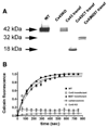
(A) Western blot showing expression of Cx43 in WT (lane 1) and in Cx43-null astrocytes transfected with full length Cx43 (lane 3), with Cx43 carboxyl terminus (lane 4) and with Cx43M257 (lane 5). Lane 2 corresponds to untransfected Cx43-null cells. Lanes 1–4 correspond to immunoblots performed with Cx43-18A antibody and lane 5 with Cx43-16A. (B) Time courses of calcein FRAP obtained from untreated (black squares) and 48 h carbenoxolone-treated (open triangles) WT astrocytes, and from untransfected (open circles) and transfected Cx43-null astrocytes with full length Cx43 (open squares) and Cx43M257 (black circles). Data were obtained from astrocytes cultured from a minimum of 3 litters of mice.
Fig. 2
Fig. 2. Modulation of P2Y1R activity by Cx43 domains
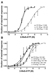
(A) Dose-response curves obtained for untreated (black squares) and 48 h carbenoxolone-treated (open triangles) WT and untransfected Cx43-null spinal cord astrocytes exposed to the P2Y1R agonist 2-MeS-ATP. Note that blockade of gap junctional communication in WT astrocytes does not alter the half-maximal response (EC50 value) induced by the P2Y1R agonist. Mean values were obtained from four to seven independent experiments. (B) Dose-response curves obtained for WT (black squares), untransfected (open circles) and transfected Cx43-null astrocytes with full length Cx43 (open squares), Cx43M257 (black circles) and Cx43CT (black triangles). Note that both full length Cx43 and Cx43CT but not Cx43M257 shifted the EC50 values of 2-MeS-ATP obtained for Cx43-null astrocytes to those obtained in WT cells. Data were obtained from 5 to 8 litters.
Fig. 3
Fig. 3. Functional interaction between P2Y1R and the SH3 domain of Cx43
(A) Dose-response curves obtained for WT (black squares) and untransfected (open circles) and Cx43Δ260–280 transfected (black triangles) Cx43-null astrocytes exposed to 2-MeS-ATP, showing that deletion of the Cx43 SH3 domain does not rescue P2Y1 receptor function. Mean±SE values are from 100 to 200 cells obtained from three litters. (B) Dose-response curves obtained from WT (black squares), and Cx43-null astrocytes untreated (open circles) and treated (black triangles) with a membrane permeant peptide corresponding to amino acids 260–280 of Cx43CT. (C) Bar histograms of the mean±SE values of intracellular calcium mobilization induced by 100 nM 2-MeS-ATP recorded from WT, Cx43-null and Cx43-null transfected with Cx43CT and Cx43M257 in the absence and presence of 5 µM PP2. Note that only untransfected and Cx43M257 transfected Cx43-null astrocytes that were not exposed to PP2 did not respond to agonist with intracellular calcium levels similar to WT astrocytes. Mean ± SE values are from 4 litters (**P < 0.001; ANOVA followed by Newman-Keuls’ Multiple Comparison Test).
Fig. 4
Fig. 4. P2Y1R expression levels in Cx43-null transfectants
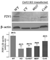
Bar histograms showing the mean values of P2Y1R expression levels in WT, untransfected Cx43-null (KO), and in Cx43-null transfected with Cx43 CT, Cx43 truncated at position 257 (M257) and with full length Cx43. An example of western blots for Cx43 and β-actin is shown above.
Fig. 5
Fig. 5. Effect of IL-1β on Cx43 and P2Y1R
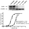
(A) Western blot showing decreased expression levels of Cx43 following exposure of spinal cord astrocytes to IL-1β (B) Dose-response curves obtained for 2-MeS-ATP performed on Fura-2 loaded WT and Cx43 KO astrocytes treated for 24 h with IL-1β (20 ng/mL). Note that exposure to the cytokine altered the agonist EC50 values in WT astrocytes. About 180 cells from three independent experiments were used in each condition.

References

    1. Ai Z, Fischer A, Spray DC, Brown AM, Fishman GI. Wnt-1 regulation of connexin43 in cardiac myocytes. J Clin Invest. 2000;105:161–171. - PMC - PubMed
    1. Ballerini P, Rathbone MP, Di Iorio P, Renzetti A, Giuliani P, D’Alimonte I, Trubiani O, Caciagli F, Ciccarelli R. Rat astroglial P2Z (P2X7) receptors regulate intracellular calcium and purine release. Neuroreport. 1996;7:2533–2537. - PubMed
    1. Beyer EC, Paul DL, Goodenough DA. Connexin43: A protein from rat heart homologous to a gap junction protein from liver. J Cell Biol. 1987;105:2621–2629. - PMC - PubMed
    1. Bezzi P, Gundersen V, Galbete JL, Seifert G, Steinhauser C, Pilati E, Volterra A. Astrocytes contain a vesicular compartment that is competent for regulated exocytosis of glutamate. Nat Neurosci. 2004;7:613–620. - PubMed
    1. Brand-Schieber E, Werner P, Iacobas DI, Iacobas S, Beelitz M, Lowery SL, Spray DC, Scemes E. Connexin43, the major gap junction protein of astrocytes, is down regulated in inflamed white matter in an animal model of multiple sclerosis. J Neurosci Res. 2005;80:798–808. - PMC - PubMed

Publication types

MeSH terms