Inhibition of VEGF expression and corneal neovascularization by siRNA targeting cytochrome P450 4B1
- PMID: 17991614
- PMCID: PMC2128778
- DOI: 10.1016/j.prostaglandins.2007.05.001
Inhibition of VEGF expression and corneal neovascularization by siRNA targeting cytochrome P450 4B1
Abstract
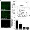
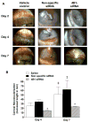
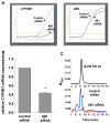
Injury to the cornea leads to formation of mediators that initiate and amplify inflammatory responses and neovascularization. Among these are lipid mediators generated by a cytochrome P450 (CYP) enzyme identified as CYP4B1. Increased corneal CYP4B1 expression increases limbal angiogenic activity through the production of 12-hydroxyeicosatrienoic acid (12-HETrE), a potent inflammatory and angiogenic eicosanoid. We used siRNA duplexes targeting CYP4B1 to substantiate the link between CYP4B1 expression, 12-HETrE production and angiogenesis in a model of suture-induced corneal neovascularization. Intrastromal sutures induced a time-dependent neovascular response which was significantly attenuated by CYP4B1-specific siRNAs but not by nonspecific siRNA. CYP4B1 mRNA was reduced by 60% and 12-HETrE's levels were barely detected in corneal homogenates from eyes treated with the CYP4B1-specific siRNA. The decreased neovascular response in CYP4B1 siRNA-treated eyes was associated with a 75% reduction in corneal VEGF mRNA levels. Transfection of rabbit corneal epithelial cells with CYP4B1 cDNA induced VEGF expression. Conversely, treatment with CYP4B1 siRNA or addition of a CYP4B1 inhibitor significantly decreased VEGF mRNA levels; addition of 12-HETrE potently increased them. The results strongly implicate the corneal CYP4B1 as a component of the inflammatory and neovascular cascade initiated by injury and further suggest that CYP4B1-12-HETrE is a proximal regulator of VEGF expression.
Figures


Similar articles
-
Corneal epithelial VEGF and cytochrome P450 4B1 expression in a rabbit model of closed eye contact lens wear.Curr Eye Res. 2001 Jul;23(1):1-10. doi: 10.1076/ceyr.23.1.1.5422. Curr Eye Res. 2001. PMID: 11821980
-
Transfection of cytochrome P4504B1 into the cornea increases angiogenic activity of the limbal vessels.J Pharmacol Exp Ther. 2005 Oct;315(1):42-50. doi: 10.1124/jpet.105.088211. Epub 2005 Jul 11. J Pharmacol Exp Ther. 2005. PMID: 16009741
-
Retinoic acid induces corneal epithelial CYP4B1 gene expression and stimulates the synthesis of inflammatory 12-hydroxyeicosanoids.J Ocul Pharmacol Ther. 2004 Feb;20(1):65-74. doi: 10.1089/108076804772745473. J Ocul Pharmacol Ther. 2004. PMID: 15006160
-
Cytochrome P450 4B1 (CYP4B1) as a target in cancer treatment.Hum Exp Toxicol. 2020 Jun;39(6):785-796. doi: 10.1177/0960327120905959. Epub 2020 Feb 13. Hum Exp Toxicol. 2020. PMID: 32054340 Review.
-
Corneal neovascularization: an anti-VEGF therapy review.Surv Ophthalmol. 2012 Sep;57(5):415-29. doi: 10.1016/j.survophthal.2012.01.007. Surv Ophthalmol. 2012. PMID: 22898649 Free PMC article. Review.
Cited by
-
Targeted suppression of HO-2 gene expression impairs the innate anti-inflammatory and repair responses of the cornea to injury.Mol Vis. 2011 Apr 29;17:1144-52. Mol Vis. 2011. PMID: 21552471 Free PMC article.
-
New Lipid Mediators in Retinal Angiogenesis and Retinopathy.Front Pharmacol. 2019 Jul 5;10:739. doi: 10.3389/fphar.2019.00739. eCollection 2019. Front Pharmacol. 2019. PMID: 31333461 Free PMC article. Review.
-
Small-interfering RNAs (siRNAs) as a promising tool for ocular therapy.Br J Pharmacol. 2013 Oct;170(4):730-47. doi: 10.1111/bph.12330. Br J Pharmacol. 2013. PMID: 23937539 Free PMC article. Review.
-
Increased expression of CYP4Z1 promotes tumor angiogenesis and growth in human breast cancer.Toxicol Appl Pharmacol. 2012 Oct 1;264(1):73-83. doi: 10.1016/j.taap.2012.07.019. Epub 2012 Jul 25. Toxicol Appl Pharmacol. 2012. PMID: 22841774 Free PMC article.
-
Heme oxygenase-1 induction attenuates corneal inflammation and accelerates wound healing after epithelial injury.Invest Ophthalmol Vis Sci. 2008 Aug;49(8):3379-86. doi: 10.1167/iovs.07-1515. Epub 2008 Apr 25. Invest Ophthalmol Vis Sci. 2008. PMID: 18441305 Free PMC article.
References
-
- Laniado Schwartzman M, Green K, Edelhauser HF, et al. Cytochrome P450 and arachidonic acid metabolism in the corneal epithelium: Role in inflammation. New York: Plenum Press; 1997.
-
- Conners MS, Stoltz RA, Webb SC, et al. A closed eye-contact lens model of corneal inflammation. I. Induction of cytochrome P450 arachidonic acid metabolism. Invest Ophthalmol Vis Sci. 1995;36:828–40. - PubMed
-
- Conners MS, Urbano F, Vafeas C, et al. Alkali burn-induced synthesis of inflammatory eicosanoids in rabbit corneal epithelium. InvestOphthalmolVis Sci. 1997;38:1963–71. - PubMed
-
- Vafeas C, Mieyal PA, Urbano F, et al. Hypoxia stimulates the synthesis of cytochrome P450-derived inflammatory eicosanoids in rabbit corneal epithelium. JPharmacolExp Ther. 1998;287:903–10. - PubMed
-
- Conners MS, Stoltz RA, Dunn MW, et al. A closed eye-contact lens model of corneal inflammation. II. Inhibition of cytochrome P450 arachidonic acid metabolism alleviates inflammatory sequelae. Invest Ophthalmol Vis Sci. 1995;36:841–50. - PubMed
Publication types
MeSH terms
Substances
Grants and funding
LinkOut - more resources
Full Text Sources

