. 2008 Feb;15(2):299-311.
doi: 10.1038/sj.cdd.4402263. Epub 2007 Nov 9.

Identification of the prosurvival activity of nerve growth factor on cardiac myocytes

Affiliations

Identification of the prosurvival activity of nerve growth factor on cardiac myocytes

A Caporali et al. Cell Death Differ. 2008 Feb.

Abstract

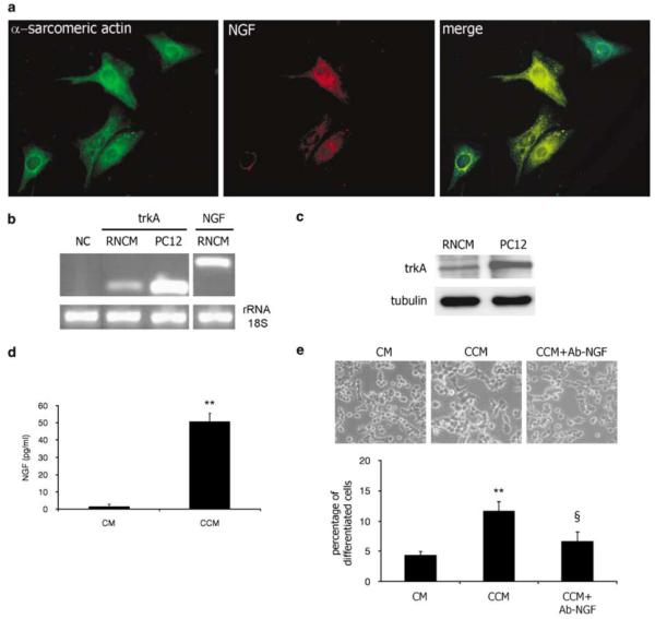
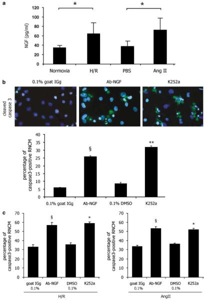
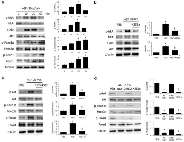
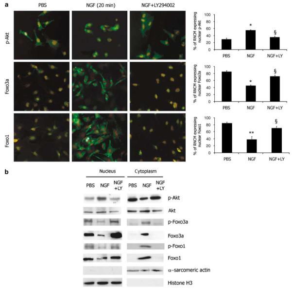
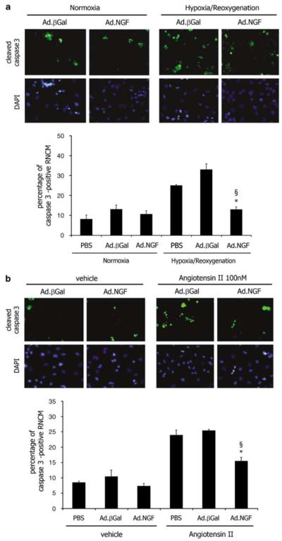
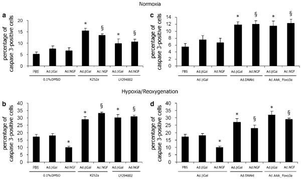
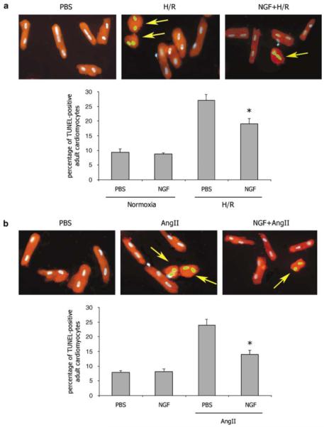
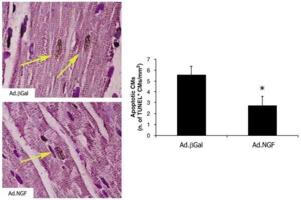
Neurotrophins (NTs) control neuron survival and regeneration. Recent research showed that NTs possess cardiovascular actions. In this study, we investigated the hypothesis that the NT nerve growth factor (NGF) prevents cardiomyocyte apoptosis. We demonstrated that cultured rat neonatal cardiomyocytes (RNCMs) produce NGF and express its trkA (tropomyosin-related receptor A (NGF high-affinity receptor)) receptor. RNCMs given a neutralizing antibody for NGF or the trkA inhibitor K252a underwent apoptosis, thus suggesting that NGF is an endogenous prosurvival factor for cardiomyocytes. Adenovirus (Ad)-mediated NGF overexpression protected RNCMs from apoptosis induced by either hypoxia/reoxygenation or angiotensin II (AngII). Similarly, recombinant NGF inhibited AngII-induced apoptosis in isolated rat adult cardiomyocytes. Finally, in a rat model of myocardial infarction, NGF gene transfer promoted cardiomyocyte survival. In RNCMs, recombinant NGF induced trkA phosphorylation, followed by Ser473 phosphorylation and nuclear translocation of phospho-protein kinase B (Akt). In response to Akt activation, Forkhead transcription factors Foxo-3a and Foxo-1 were phosphorylated and excluded from the nucleus. The prosurvival effect of adenoviral vector carrying the human NGF gene was inhibited in vitro by K252a, LY294002 (a pan-phosphatidyl inositol 3-kinase - PI3K - inhibitor), an Akt small interfering RNA, and adenoviruses carrying a dominant negative mutant form of Akt (Ad.DN.Akt) or an Akt-resistant Foxo-3a (Ad.AAA-Foxo-3a). These results newly demonstrate the cardiac prosurvival action of NGF and provide mechanistic information on the signaling pathway, which encompasses trkA, PI3K-Akt, and Foxo.


Figures

Figure 1
Figure 1
Rat neonatal cardiomyocytes (RNMCs) express NGF and trkA. (a) Immunofluorescence analysis of the cardiac marker α-sarcomeric actin (green fluorescence) and NGF (red fluorescence) and merged images (costaining results in yellow fluorescence). (b) RT-PCR analysis of trkA and NGF expression in RNMCs. PC12 cells, which express high level of trkA, were used for reference. Bands are representative of three experiments. Ribosomal RNA (18S) was used as a loading control. Water was used as negative control (NC). (c) Western blot analysis of trkA in lysates of RNCMs. PC12 protein extracts were loaded as positive controls for trkA. Bands are representative of three experiments. (d) Determination of NGF content in the RNCM conditioned culture medium (CCM) and in basal culture medium (CM) by ELISA. Data are presented as mean±S.E.M. (n = 3). **P<0.01 versus CM. (e) Secreted NGF from RNCM-mediated differentiation (neurite extension) of PC12 cells. PC12 morphology was observed after incubation for 3 days in the CCM of RNCMs or in CM. A NGF-neutralizing antibody (Ab-NGF) prevented PC12 differentiation. Data are presented as mean±S.E.M. (n=3). **P<0.01 versus CM; §P<0.05 versus CCM
Figure 2
Figure 2
The release of NGF from RNCMs increases under proapoptotic conditions and NGF is an endogenous prosurvival factor for RNCMs. (a) NGF content in the serum-free culture of RNCMs exposed to hypoxia/reoxygenation (H/R, control: normoxia) or angiotensin II (AngII, control: PBS) was measured by ELISA. Respective controls were the serum-free conditioned medium of normoxic RNCMs and PBS-treated RNCMs, respectively. Data are presented as mean±S.E.M. (n = 3). *P<0.05 versus the respective control. (b) RNCMs were incubated for 48 h in serum-free medium with Ab-NGF (raised in goat) or the trkA inhibitor K252a. Controls consisted of non-immune goat serum (0.1% in PBS) or 0.1% DMSO, respectively. Apoptosis was detected by cleaved caspase-3 immuno-staining (green fluorescence). Nuclei were counterstained by DAPI (blue fluorescence). The fluorescent images (× 400) are representative of the experiment. Bar graph shows the percentage of cleaved caspase 3-positive RNCMs. Data are presented as mean±S.E.M. (n = 3). §P<0.05 versus 0.1% goat IgG; **P<0.01 versus 0.1% DMSO. (c) RNCMs were submitted to H/R or incubated with AngII (for both as described in (a)) in the presence of Ab-NGF, K252a, or the respective controls. Apoptosis was evaluated by cleaved caspase-3 staining, as described in (b). Data are presented as mean±S.E.M. (n = 3). §P<0.05 versus 0.1% goat IgG; *P<0.05 versus 0.1% DMSO
Figure 3
Figure 3
NGF signaling in RNMCs. (a) Serum-starved RNCMs received NGF (50 ng/ml). After 0, 10, 20, and 60 min, the phosphorylation of trkA (Tyr490), Akt (Ser473), Foxo-3a (Thr32), and Foxo-1 (Ser256) was determined by western blot. Tubulin was used as loading control. Graphs show the ratio between the densitometric reading of phosphorylated and total trkA, Akt, Foxo-3a, and Foxo-1. Data are presented as mean±S.E.M. (n=3). * P<0.05 versus time zero. (b) Serum-starved RNCMs were pretreated with the trkA inhibitor K252a (100 nM) or its vehicle (0.1% DMSO) before adding NGF. After 20 min, the phosphorylation of trkA and Akt was examined. Controls (PBS) received 0.1% DMSO (instead of K252a) and PBS (instead of NGF). Tubulin was used as loading control. K252a blocked the phosphorylation of both trkA and Akt. Graphs show the ratio between the densitometric reading of phosphorylated and total trkA and phosphorylated and total Akt. Data are presented as mean±S.E.M. (n=3). *P<0.05 versus PBS; §P<0.05 versus NGF + 0.1% DMSO (NGF). (c) Serum-starved RNMCs were pretreated with the PI3K inhibitor LY294002 (50 μM) or its vehicle (0.1% DMSO) before adding NGF. After 20 min, the phosphorylation of Akt, Foxo-3a, and Foxo-1 was evaluated. Controls received 0.1% DMSO and PBS. Tubulin was used as loading control. LY294002 blocked the phosphorylation of Akt, Foxo-3a, and Foxo-1. Graphs show the ratio between the densitometric reading of phosphorylated and total Akt, phosphorylated and total Foxo-3a, and phosphorylated and total Foxo-1. Data are presented as mean±S.E.M. (n=3). *P<0.05 versus PBS; §P<0.05 versus NGF + 0.1% DMSO (NGF). (d) Serum-free-cultured RNCMs were treated for 48 h with Ab-NGF or K252a. Controls received 0.1% nonimmune goat serum or 0.1% DMSO. Tubulin was used as loading control. Either Ab-NGF or K252a reduced phosphorylation of Akt, Foxo-3a, and Foxo-1. Graphs show the ratio between the densitometric reading of phosphorylated and total Akt, phosphorylated and total Foxo-3a, and phosphorylated and total Foxo-1. Data are presented as mean±S.E.M. (n 3). *P<0.05 versus 0.1% goat IgG; §P<0.01 versus 0.1% DMSO
Figure 4
Figure 4
NGF induces nuclear/cytoplasmatic shuttling of phospho-Akt and Foxo-3a and Foxo-1 in RNCMs. (a) Serum-starved RNCMs were pretreated with LY294002 or its vehicle (0.1% DMSO) before receiving NGF (50 ng/ml) or PBS. After 20 min, cells were fixed and stained for phospho-Akt, total Foxo-3a, or total Foxo-1 by using FITC-conjugated antibodies (green fluorescence). Nuclei were stained by PI (red fluorescence). Exposure was optimized to demonstrate localization and it does not reflect the concentration of the antigen. NGF induced nuclear localization of phospho-Akt (as demonstrated by the yellow/orange colors of nuclei, which indicates colocalization of FITC and PI) and promoted nuclear export of Foxo-3a and Foxo-1. LY294002 abolished these NGF effects. Graphs show the percentage of RNCMs expressing nuclear phospho-Akt, Foxo-3a, and Foxo-1. Results are from three independent experiments. Data are presented as mean±S.E.M. (n = 3). *P<0.05 and **P<0.01 versus PBS; §P<0.05 versus NGF + 0.1% DMSO (NGF). (b) Serum-starved RNCMs were pretreated with LY294002 or its vehicle (0.1% DMSO) for 30 min before receiving NGF (50 ng/ml) or PBS. After 20 min, cells were lysated and nuclear and cytoplasmatic fraction prepared. Nuclear and cytoplasmatic extracts were assessed by western blot using antibodies targeting phospho- and total form of Akt, Foxo-3a, and Foxo-1. Efficient separation of nuclear and cytoplasmatic fractions was confirmed by western blot for histone H3 and α-sarcomeric actin, respectively
Figure 5
Figure 5
NGF overexpression prevents apoptosis of RNCMs. RNCMs were transduced with Ad.NGF or Ad.βGal, or not infected (PBS). (a) RNCMs underwent 12 h of hypoxia followed by 24 h reoxygenation or were maintained under normoxia. (b) RNCMs were infected with Ad.NGF or Ad.βGal or given PBS before being stimulated with AngII (10−7 M, for 24 h). Apoptotic RNCMs were detected by cleaved caspase-3 staining (green fluorescence). Bar graphs show the quantitative analysis of apoptosis, which is expressed as percentage of cleaved caspase-3-positive RNCMs. Data are presented as mean±S.E.M. (n = 3). *P<0.05 versus PBS; §P<0.05 versus Ad.βGal. Representative images are presented above graphs
Figure 6
Figure 6
NGF overexpression prevents apoptosis of RNCMs through trkA, Akt, and Foxo-3a. (a, b) RNCMs were infected with Ad.NGF or AdβGal and then given K252a, LY294002, or 0.1% DMSO (vehicle). Not infected RNCMs were given PBS and 0.1% DMSO were used for reference. (c, d) RNCMs were coinfected with Ad.NGF or Ad.βGal (as control of Ad.NGF) and with an adenovirus carrying a dominant negative mutant form of Akt (Ad.DN-Akt), an adenovirus carrying a constitutionally active form of Foxo-3a (Ad.AAA-Foxo-3a), or Ad.βGal (as control of both Ad.DN-Akt and Ad.AAA-Foxo-3a. RNCMs infected with Ad.βGal and given PBS were used for reference. RNCMs in (a) and (c) were maintained under normoxia. RNCMs in (b) and (d) underwent 5 h hypoxia and 24 h reoxygenation. Apoptosis was revealed by cleaved caspase-3 staining. Graphs show the quantitative analysis of apoptosis, which is expressed as percentage of cleaved caspase-3-positive RNCMs. Data are presented as mean±S.E.M. (n=3). (a, b): *P<0.05 versus Ad.βGal plus 0.1% DMSO; §P<0.05 versus Ad.NGF plus 0.1% DMSO. (c, d): *P<0.05 versus Ad.βGal plus Ad.βGal; §P<0.05 versus Ad.NGF plus Ad.βGal
Figure 7
Figure 7
NGF inhibits apoptosis in rat adult cardiomycytes. (a) Isolated rat adult cardiomyocytes were maintained under normoxia or submitted to 6 h hypoxia followed by 18 h reoxygenation and cotreated with NGF (50 ng/ml) or its vehicle PBS. (b) Rat adult cardiomyocytes were incubated with AngII (10−9 M) or its vehicle PBS and cotreated with NGF (50 ng/ml) or its vehicle PBS for 24 h. Apoptotic nuclei were identified by TUNEL staining (green fluorescence). α-Sarcomeric actin stains cardiomyocytes (red fluorescence). The pictures were captured at ×400magnification. Arrows point to TUNEL-positive cardiomyocytes. Bar graphs quantify apoptosis, which is expressed as percentage of TUNEL-positive cardiomyocyte. Data are presented as mean±S.E.M. (n = 3). (a): *P<0.05 versus PBS plus H/R; (b): *P<0.05 versus PBS plus AngII
Figure 8
Figure 8
Local NGF gene transfer prevents apoptosis of cardiomyocytes in the rat infarcted heart. Myocardial infarction (MI) was induced in adult Wistar rat. Ad.NGF or Ad.βGal (each at 108 p.f.u.) was injected in the peri-infarct myocardium. After 7 days, the heart was arrested in diastole and perfusion/fixed. Apoptosis of cardiomyocytes (CMs) was revealed by double staining for TUNEL (TUNEL-positive nuclei are stained in dark brown) and the cardiac marker α-sarcomeric actin (in purple). Nuclei were counterstained with hematoxylin. In the pictures captured (optical microscopy, ×1000) from Ad.NGF and Ad.βGal specimens, TUNEL-positive apoptotic cardiomyocytes are pointed by arrows. Graph quantifies apoptosis of cardiomyocytes per mm2 of peri-infarct myocardium section. Values are mean±S.E.M. (n = 7) *P<0.05 versus Ad.βGal

Similar articles

Cited by

References

    1. Levi-Montalcini R. The nerve growth factor: thirty-five years later. EMBO J. 1987;6:1145–1154. - PMC - PubMed
    1. Chao MV, Rajagopal R, Lee FS. Neurotrophin signalling in health and disease. Clin Sci (Lond) 2006;110:167–173. - PubMed
    1. Emanueli C, Salis MB, Pinna A, Graiani G, Manni L, Madeddu P. Nerve growth factor promotes angiogenesis and arteriogenesis in ischemic hindlimbs. Circulation. 2002;106:2257–2262. - PubMed
    1. Cantarella G, Lempeur L, Presta M, Ribatti D, Lombardo G, Lazarovici P, et al. Nerve growth factor–endothelial cell interaction leads to angiogenesis in vitro and in vivo. FASEB J. 2002;16:1307–1309. - PubMed
    1. Salis MB, Graiani G, Desortes E, Caldwell RB, Madeddu P, Emanueli C. Nerve growth factor supplementation reverses the impairment, induced by type 1 diabetes, of hindlimb post-ischaemic recovery in mice. Diabetologia. 2004;47:1055–1063. - PubMed

Publication types

MeSH terms