. 2007 Dec;117(12):3833-45.
doi: 10.1172/JCI32567.

TNF-alpha is critical for antitumor but not antiviral T cell immunity in mice

Affiliations

TNF-alpha is critical for antitumor but not antiviral T cell immunity in mice

Thomas Calzascia et al. J Clin Invest. 2007 Dec.

Abstract

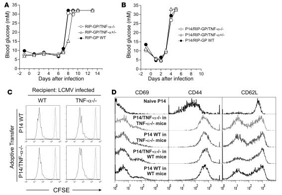
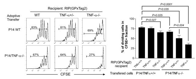
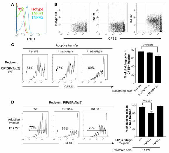
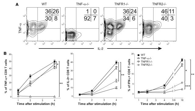
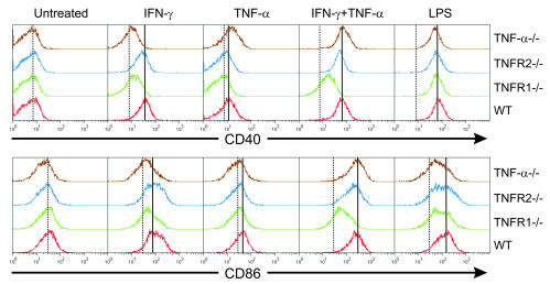
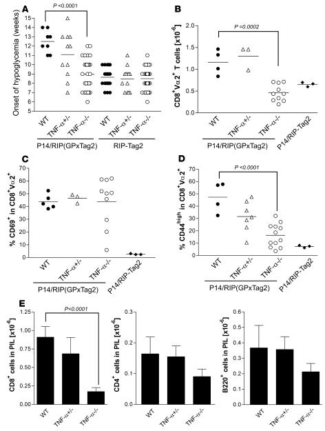
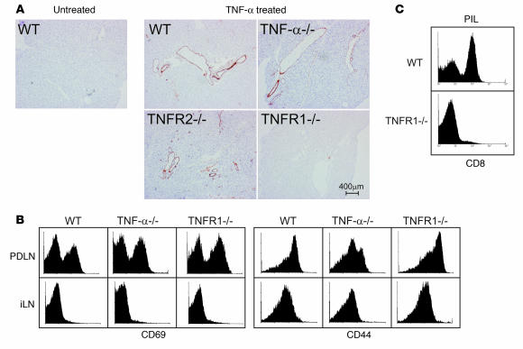
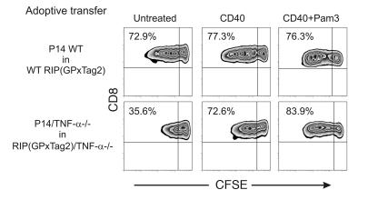
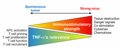
TNF-alpha antagonists are widely used in the treatment of inflammatory and autoimmune diseases, but their use is associated with reactivation of latent infections. This highlights the importance of TNF-alpha in immunity to certain pathogens and raises concerns that critical aspects of immune function are impaired in its absence. Unfortunately, the role of TNF-alpha in the regulation of T cell responses is clouded by a myriad of contradictory reports. Here, we show a role for TNF-alpha and its receptors, TNFR1 and TNFR2, specifically in antitumor immunity. TNF-alpha-deficient mice exhibited normal antiviral responses associated with strong inflammation. However, TNF-alpha/TNFR1-mediated signals on APCs and TNF-alpha/TNFR2 signals on T cells were critically required for effective priming, proliferation, and recruitment of tumor-specific T cells. Furthermore, in the absence of TNF-alpha signaling, tumor immune surveillance was severely abrogated. Finally, treatment with a CD40 agonist alone or in combination with TLR2 stimuli was able to rescue proliferation of TNF-alpha-deficient T cells. Therefore, TNF-alpha signaling may be required only for immune responses in conditions of limited immunostimulatory capacity, such as tumor surveillance. Importantly, these results suggest that prolonged continuous TNF-alpha blockade in patients may have long-term complications, including potential tumor development or progression.


Figures

Figure 1
Figure 1. The absence of TNF-α does not alter the kinetics or magnitude of LCMV-induced autoimmune diabetes in RIP-GP or P14/RIP-GP animals.
(A) RIP-GP mice were infected with LCMV Arm. Blood glucose levels were measured at multiple time points and used as an indication of islet β cell autoimmune destruction. Blood glucose levels of 1 mouse representative of at least 6 mice are shown for each genotype. (B) P14/RIP-GP animals were infected with LCMV Arm, and blood glucose was monitored as in A. Blood glucose levels of 1 mouse representative of at least 5 mice are shown for each genotype. (C) CFSE-labeled P14 WT or TNF-α–/– CD8+ T cells were adoptively transferred into WT or TNF-α–/– mice infected 3 days previously with LCMV Arm. The extent of T cell proliferation was assessed by CFSE dilution in spleens 3 days later (solid lines). Histograms are gated on CFSE+CD8+Vα2+ cells and are representative of 3 independent animals per condition. Dashed lines indicate the CFSE profile of undivided cells. (D) Expression of CD69, CD44, and CD62L on CFSE+CD8+Vα2+ cells from C.
Figure 2
Figure 2. Intrinsic and extrinsic defects impair T cell proliferation in response to spontaneous malignancies in TNF-α–/– mice.
CFSE-labeled naive P14 WT or P14/TNF-α–/– CD8+ T cells were adoptively transferred into the indicated RIP(GP × Tag2) hosts. Proliferation of CFSE-labeled cells was assessed 3 days later in PDLN. Histograms are gated on CFSE+CD8+Vα2+ cells. The percentage in each histogram indicates the proportion of dividing cells among the CFSE-labeled population. A summary and mean of the proportion of proliferating cells observed in individual animals is presented. Error bars indicate SEM The indicated differences are statistically significant. Number of mice analyzed: WT→WT: n = 6; WT→TNF-α+/–: n = 3; WT→TNF-α–/–: n = 6; TNF-α–/–→WT: n = 4; TNF-α–/–→TNF-α+/–: n = 3; and TNF-α–/–→TNF-α–/–: n = 5.
Figure 3
Figure 3. TNFR2 but not TNFR1 expression on T cells integrates TNF-α–dependent costimulatory signals.
(A) TNFR1 and TNFR2 expression on naive P14 T cells was assessed by gating on CD8+Vα2+ cells in inguinal LN (iLN) of P14/RIP(GP × Tag2) animals. (B) Proliferating P14 cells display high levels of TNFR2 expression. CFSE-labeled naive P14 WT CD8+ T cells were adoptively transferred into WT RIP(GP × Tag2) hosts. TNFR1 and TNFR2 modulation during proliferation was assessed 3 days later in PDLN. Dot plots are gated on CFSE+CD8+Vα2+ cells. The data shown are representative of 3 independent experiments. (C) CFSE-labeled naive P14 WT, P14/TNFR1–/–, or P14/TNFR2–/– CD8+ T cells were adoptively transferred into WT RIP(GP × Tag2) hosts. Proliferation of CFSE-labeled cells was assessed 3 days later in PDLN. Histograms are gated on CFSE+CD8+Vα2+ cells. The percentage in each histogram indicates the proportion of dividing cells among the CFSE-labeled population. A summary and mean of the proportion of proliferating cells observed in individual animals is presented. Error bars indicate SEM. Number of mice analyzed: P14 WT: n = 7; P14/TNFR1–/–: n = 6; P14/TNFR2–/–: n = 7. (D) CFSE-labeled naive P14 WT CD8+ T cells were adoptively transferred into WT, TNFR1–/–, or TNFR2–/– RIP(GP × Tag2) recipients of the indicated genotype. Proliferation of CFSE-labeled cells was assessed 3 days later in PDLN. Histograms were gated on CFSE+CD8+Vα2+ cells. A summary and mean of the proportion of proliferating cells observed in individual animals is presented. Error bars indicate SEM. A significant reduction in proliferation was observed in TNFR1–/– RIP(GP × Tag2) mice (P = 0.037). Number of mice analyzed: WT RIP(GP × Tag2): n = 5; RIP(GP × Tag2)/TNFR1–/–: n = 8; RIP(GP × Tag2)/TNFR2–/–: n = 3.
Figure 4
Figure 4. TNF-α/TNFR2–related T cell–intrinsic defects result in defective cytokine production.
(A) P14 WT, P14/TNFR1–/–, P14/TNFR2–/–, or P14/TNF-α–/– CD8+ T cells were cocultured with GP33-pulsed thioglycollate-induced macrophages for 4 hours in the presence of an inhibitor of intracellular protein transport. Following incubation, P14 cells were harvested, and the levels of intracellular TNF-α, IL-2, and IFN-γ were evaluated by intracellular flow cytometry. Representative contour plots for IL-2 and TNF-α are shown. The proportion of cells in each quadrant is also indicated. (B) Kinetics of TNF-α, IL-2, and IFN-γ production by P14 WT, P14/TNFR1–/–, P14/TNFR2–/–, or P14/TNF-α–/– CD8+ T cells stimulated as described in A. Mean values ± SEM from 4 independent experiments are shown. Statistically significant differences at 4 hours are indicated. For TNF-α, *P = 0.0249, **P = 0.0001. For IL-2, *P = 0.0048, **P = 0.0273. For IFN-γ, *P = 0.0122, **P < 0.0001, ***P = 0.0003.
Figure 5
Figure 5. TNF-α and TNFR1 on APCs are required for optimal activation in response to inflammatory cytokines.
Bone marrow–derived macrophages were generated from mice of the indicated genotypes and treated for 20 hours with IFN-γ, TNF-α, or LPS. The levels of CD40 and CD86 expression were then evaluated by flow cytometry. Data are representative of 2 independent experiments.
Figure 6
Figure 6. Reduced T cell expansion and tumor infiltration in P14/RIP(GP × Tag2)/TNF-α–/– animals.
(A) Impaired immune surveillance in TNF-α–/– P14/RIP(GP × Tag2) animals. The age at onset of hypoglycemia was determined by weekly measurement of blood glucose levels in mice of the indicated genotype. This was used as a surrogate marker of tumor development and immune surveillance, since it is a direct measure of β islet cell mass. Results obtained for individual mice are shown. Differences between WT and TNF-α–/– P14/RIP(GP × Tag2) animals were statistically significant (P < 0.0001). (B) The number of CD8+Vα2+ P14 cells in PDLN of P14/RIP(GP × Tag2) animals of the indicated genotypes was calculated after flow cytometry analysis of PDLN. Averages and data obtained from individual mice are shown. Differences between WT and TNF-α–/– animals were statistically significant (P = 0.0002). (C and D) The proportion of GP-specific P14 CD8+ T cells expressing high levels of the activation markers CD69 (C) and CD44 (D) was assessed in PDLN after gating on CD8+Vα2+ cells. Averages and data obtained from individual mice are shown. iLN data are also shown as a control. Differences between WT and TNF-α–/– animals were statistically significant for CD44 (P < 0.0001). (E) Pancreas-infiltrating leukocytes (PIL) were isolated from P14/RIP(GP × Tag2) WT, TNF-α+/–, or TNF-α–/– pancreas, and the number of infiltrating CD8+, CD4+, and B220+ cells was determined by flow cytometry and analysis of the leukocytic infiltrates. Error bars indicate SEM. The number of CD8+ cells in PILs from P14/RIP(GP × Tag2)/TNF-α–/– mice was significantly lower than in those from P14 WT/RIP(GP × Tag2) animals (P < 0.0001) (WT mice: n = 5; TNF-α–/– mice: n = 10).
Figure 7
Figure 7. TNFR1 signaling modulates the expression of adhesion molecules on pancreatic vessels and favors recruitment of GP-specific CD8+ T cells to the tumor site.
(A) Histological sections of pancreas from mice of the indicated genotypes were stained for VCAM-1 7 hours following i.p. injection of recombinant mouse TNF-α. (B) T cell activation in PDLN of P14/RIP(GP × Tag2)/TNFR1–/– animals. Comparison of the expression of CD69 and CD44 on CD8+Vα2+ cells from PDLN or iLN of WT, TNF-α–/–, or TNFR1–/– P14/RIP(GP × Tag2) animals. Representative results from at least 4 independent experiments on different mice are shown. (C) Impaired CD8+ T cell recruitment to pancreas of P14/RIP(GP × Tag2)/TNFR1–/– animals. Proportion of CD8+ T cells in PILs from WT or TNFR1–/– animals. A representative result from at least 4 independent mice is shown.
Figure 8
Figure 8. The exogenous administration of immunostimulatory factors rescues the proliferation of P14/TNF-α–/– T cells in PDLN of RIP(GP × Tag2)/TNF-α–/– mice.
CFSE-labeled naive P14 WT or P14/TNF-α–/– CD8+ T cells were adoptively transferred into the indicated RIP(GP × Tag2) hosts in combination with CD40 agonistic Ab or CD40 Ab and the TLR2 ligand Pam3. Proliferation of CFSE-labeled cells was assessed 3 days later in PDLN. Zebra plots were gated on CFSE+CD8+Vα2+ cells. The percentage in each plot indicates the proportion of dividing cells among the CFSE-labeled population. The data presented are representative of 3 independent experiments.
Figure 9
Figure 9. Model depicting the inverse association between immunostimulatory strength and inflammation and the requirement for TNF-α in T cell immune responses.
In conditions when T cells are activated in the presence of optimal stimulation (optimal APC maturation by danger signals, costimulation, cytokines, etc.), TNF-α is totally or partially dispensable for T cell activation (e.g., systemic viral infection), while when such factors become limiting (e.g., spontaneous tumor growth), TNF-α’s roles become relevant and critical at multiple levels of the T cell immune response.

Similar articles

Cited by

References

    1. Carswell E.A., et al. An endotoxin-induced serum factor that causes necrosis of tumors. Proc. Natl. Acad. Sci. U. S. A. 1975;72:3666–3670. - PMC - PubMed
    1. MacEwan D.J. TNF receptor subtype signalling: differences and cellular consequences. Cell Signal. 2002;14:477–492. - PubMed
    1. Wajant H., Pfizenmaier K., Scheurich P. Tumor necrosis factor signaling. Cell Death Differ. 2003;10:45–65. - PubMed
    1. McDevitt H., Munson S., Ettinger R., Wu A. Multiple roles for tumor necrosis factor-α and lymphotoxin α/β in immunity and autoimmunity. Arthritis Res. 2002;4(Suppl. 3):S141–S152. - PMC - PubMed
    1. Kollias G. TNF pathophysiology in murine models of chronic inflammation and autoimmunity. Semin. Arthritis Rheum. 2005;34:3–6. - PubMed

Publication types

MeSH terms

Substances