. 2007 Dec;117(12):3664-72.
doi: 10.1172/JCI28115.

Human epithelial cells establish direct antifungal defense through TLR4-mediated signaling

Affiliations

Human epithelial cells establish direct antifungal defense through TLR4-mediated signaling

Günther Weindl et al. J Clin Invest. 2007 Dec.

Abstract

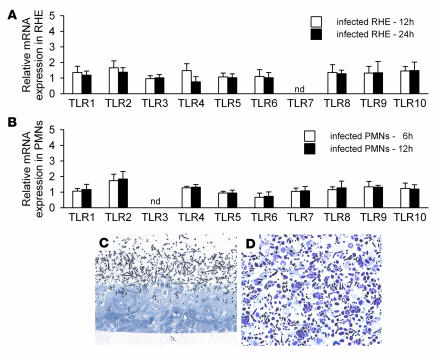
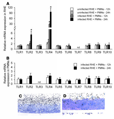
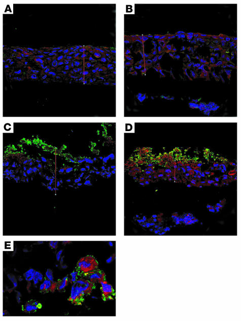
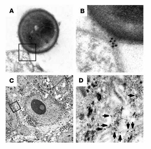
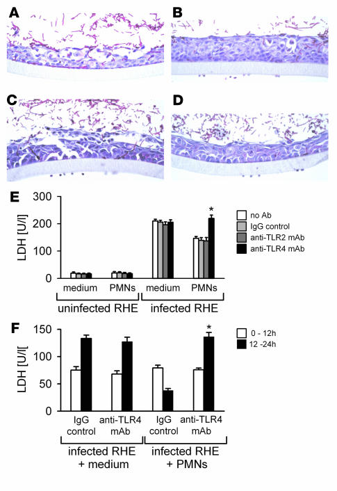
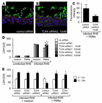
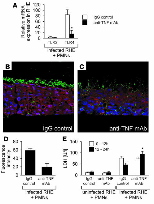
Mammalian TLRs are central mediators of the innate immune system that instruct cells of the innate and adaptive response to clear microbial infections. Here, we demonstrate that human epithelial TLR4 directly protected the oral mucosa from fungal infection via a process mediated by polymorphonuclear leukocytes (PMNs). In an in vitro epithelial model of oral candidiasis, the fungal pathogen Candida albicans induced a chemoattractive and proinflammatory cytokine response but failed to directly modulate the expression of genes encoding TLRs. However, the addition of PMNs to the C. albicans-infected model strongly upregulated cytoplasmic and cell-surface epithelial TLR4 expression, which correlated directly with protection against fungal invasion and cell injury. C. albicans invasion and cell injury was restored by the addition of TLR4-specific neutralizing antibodies and knockdown of TLR4 using RNA interference, even in the presence of PMNs, demonstrating the direct role of epithelial TLR4 in the protective process. Furthermore, treatment with neutralizing antibodies specific for TNF-alpha resulted in strongly reduced TLR4 expression accompanied by augmented epithelial cell damage and fungal invasion. To our knowledge, this is the first description of such a PMN-dependent, TLR4-mediated protective mechanism at epithelial surfaces, which may provide significant insights into how microbial infections are managed and controlled in the oral mucosa.


Figures

Figure 1
Figure 1. Interaction of oral RHE and PMNs with C. albicans does not alter the constitutive levels of TLR1-10 expression.
(A and B) TLR1–TLR10 mRNA levels in C. albicans–infected RHE (A, n = 6) and after coincubation of PMNs with C. albicans (B, n = 6). Values are normalized to housekeeping genes YWHAZ and G6PD and relative to uninfected RHE or PMNs (control, assigned as 1.0). nd, not detected. (C and D) Light micrographs of oral RHE 24 h after C. albicans infection (C) and of PMNs 12 h after coincubation with C. albicans (D). Original magnification, ×40.
Figure 2
Figure 2. PMNs induce epithelial TLR4 gene upregulation, which protects against fungal invasion and cell damage, in the presence of C. albicans.
(A) TLR expression in uninfected and infected oral RHE in the presence of PMNs. Expression values are normalized to YWHAZ and G6PD and relative to uninfected RHE without PMNs (control, assigned as 1.0). *P < 0.05, 2-tailed paired Student’s t test. (B) TLR expression in the PMNs isolated from the filter of the C. albicans–infected RHE, relative to PMNs isolated from uninfected RHE (control, assigned as 1.0) and normalized to YWHAZ (n = 6). (C and D) C. albicans–infected oral RHE after 24 h. In the absence of PMNs, extensive edema and vacuolization was observed (C), while a strongly reduced fungal virulence phenotype was evident following PMN addition to the basal side of the filter after 12 h (D). Original magnification, ×40.
Figure 3
Figure 3. PMNs stimulate TLR4 protein expression in epithelial cells in direct contact with C. albicans.
Confocal laser microscopy of oral RHE after 24 h in the presence and absence of C. albicans and PMNs (cell nuclei, blue; TLR4, red; C. albicans, green). Basal expression (A) of TLR4 in the oral RHE was nearly unchanged after the addition of PMNs alone (B) or C. albicans alone (C). (D) Strong upregulation of TLR4 was clearly evident in the C. albicans–infected oral RHE in the presence of PMNs. (E) Higher-magnification image demonstrating increased TLR4 expression only in epithelial cells in direct contact with C. albicans in the presence of PMNs. Data are representative of 3 independent experiments. Original magnification, ×40 (AD); ×250 (E).
Figure 4
Figure 4. Cell surface TLR4 directly recognizes C. albicans and is expressed intracellularly.
Electron microscopy with postembedding immunogold labeling in C. albicans–infected oral RHE at 24 h in the presence of PMNs. (A) Expression of TLR4 was evident at the site of direct host-pathogen contact. (B) Higher-magnification view of boxed region in A. (C) An epithelial cell inside the oral RHE. (D) Higher-magnification view of boxed region in C revealed expression of TLR4 at the cell membrane and intracellularly. Arrows indicate gold-labeled TLR4 expression. Data are representative of 3 independent experiments. Original magnification, ×20,000 (A and B); ×3,000 (C and D).
Figure 5
Figure 5. TLR4 blocking antibody reverses the antifungal protective effect of PMNs on the oral mucosa.
(AD) Light micrographs of RHE 24 h after infection with C. albicans SC5314 in the absence and presence of PMNs. (A) Invasion of C. albicans with extensive edema and vacuolization of all epithelial layers in the absence of PMNs. (B) Strongly reduced virulence phenotype of C. albicans at the same time point when PMNs were added after 12 h to the basal side of the filter. (C) In the presence of anti-TLR4 neutralizing mAbs, C. albicans–induced tissue damage was restored, while (D) isotype-matched control IgG antibodies showed no effect on C. albicans virulence. PMNs were attached to the basal side of the filter in BD. Data are representative of 3 experiments. Original magnification, ×40. (E) Release of LDH by epithelial cells 24 h after infection (or not) with C. albicans in the presence and absence of PMNs (added after 12 h) (n = 6). (F) LDH values before (0–12 h) and after (12–24 h) addition of PMNs to C. albicans–infected RHE model (n = 4). *P < 0.05, 2-tailed paired Student’s t test.
Figure 6
Figure 6. Knockdown of epithelial TLR4 by siRNA abrogates the protective phenotype.
(A and B) Confocal microscopy of TLR4 expression in the RHE. C. albicans infection of the RHE and supplementation with PMNs showed strong epithelial TLR4 expression 24 h after transfection with control siRNA (A), while decreased TLR4 expression associated with increased fungal invasion was evident after transfection with 10 nM TLR4 siRNA2 (B). Original magnification, ×40. (C) Mean fluorescence intensity of TLR4 confirmed reduced expression. Data are representative of 3 independent experiments. (D) Release of LDH by epithelial cells 24 h after infection (or not) with C. albicans in the presence and absence of PMNs (added after 12 h) (n = 6). (E) LDH values before (0–12 h) and after (12–24 h) addition of PMNs to C. albicans–infected RHE model (n = 4). *P < 0.05, 2-tailed paired Student’s t test.
Figure 7
Figure 7. Neutralization of TNF-α release reduces TLR4 expression and restores fungal invasion.
(A) TLR4 mRNA expression was strongly decreased after incubation of the oral RHE and PMNs with anti–TNF-α neutralizing mAbs. Values are normalized to YWHAZ and G6PD and relative to uninfected RHE without PMNs (control, assigned as 1.0). (B and C) Confocal microscopy of TLR4 expression in the infected RHE in the presence of PMNs showed prominent epithelial TLR4 expression at 24 h after treatment with IgG control antibody (B), while decreased TLR4 expression and increased fungal invasion was evident after incubation with anti–TNF-α mAbs, particularly in the basal side of the epithelium (C). Original magnification, ×40. (D) Reduced mean fluorescence intensity was observed after neutralization of TNF-α. Data are representative of 3 independent experiments. (E) Release of LDH by epithelial cells 24 h after infection (or not) with C. albicans in the presence of PMNs (added after 12 h). LDH values before (0–12 h) and after (12–24 h) addition of PMNs to C. albicans–infected RHE model (n = 3). *P < 0.05, 2-tailed paired Student’s t test.

Similar articles

Cited by

References

    1. Godaly G., et al. Neutrophil recruitment, chemokine receptors, and resistance to mucosal infection. J. Leukoc. Biol. 2001;69:899–906. - PubMed
    1. Backhed F., Hornef M. Toll-like receptor 4-mediated signaling by epithelial surfaces: necessity or threat? Microbes Infect. 2003;5:951–959. - PubMed
    1. Tjabringa G.S., et al. Host defense effector molecules in mucosal secretions. FEMS. Immunol. Med. Microbiol. 2005;45:151–158. - PubMed
    1. Takeda K., Kaisho T., Akira S. Toll-like receptors. Annu. Rev. Immunol. 2003;21:335–376. - PubMed
    1. Roeder A., Kirschning C.J., Rupec R.A., Schaller M., Korting H.C. Toll-like receptors and innate antifungal responses. Trends Microbiol. 2004;12:44–49. - PubMed

Publication types

MeSH terms